
Definition
Tibialis anterior tendinopathy is a painful condition of the tendon of the tibialis anterior muscle, usually related to gradual mechanical overload of this tendon, which lies on the front of the ankle. It mainly presents as pain on the front of the ankle, along the course of the tendon, sometimes on the medial border of the midfoot, and particularly during dorsiflexion movements of the foot (pulling the toes up towards you).
It is a relatively rare condition compared with Achilles or tibialis posterior tendinopathy. In athletes, it most often appears after a substantial increase in running volume, an increase in elevation gain, hill work, a change of shoes, a change in running technique or a return after a period off.
It is essential to distinguish tendinopathy from tenosynovitis, a partial tear or a rupture of the tibialis anterior tendon — the latter being a different clinical situation, which may require specific surgical management (see the Surgery section).
Anatomy
The tibialis anterior lies in the anterior compartment of the leg. It originates mainly from the lateral surface of the tibia, the interosseous membrane and the adjacent fascial structures. Its tendon runs down the front of the ankle, then passes beneath the extensor retinaculum before usually inserting on the medial cuneiform and the base of the first metatarsal — although there are anatomical variations in its distal insertion.
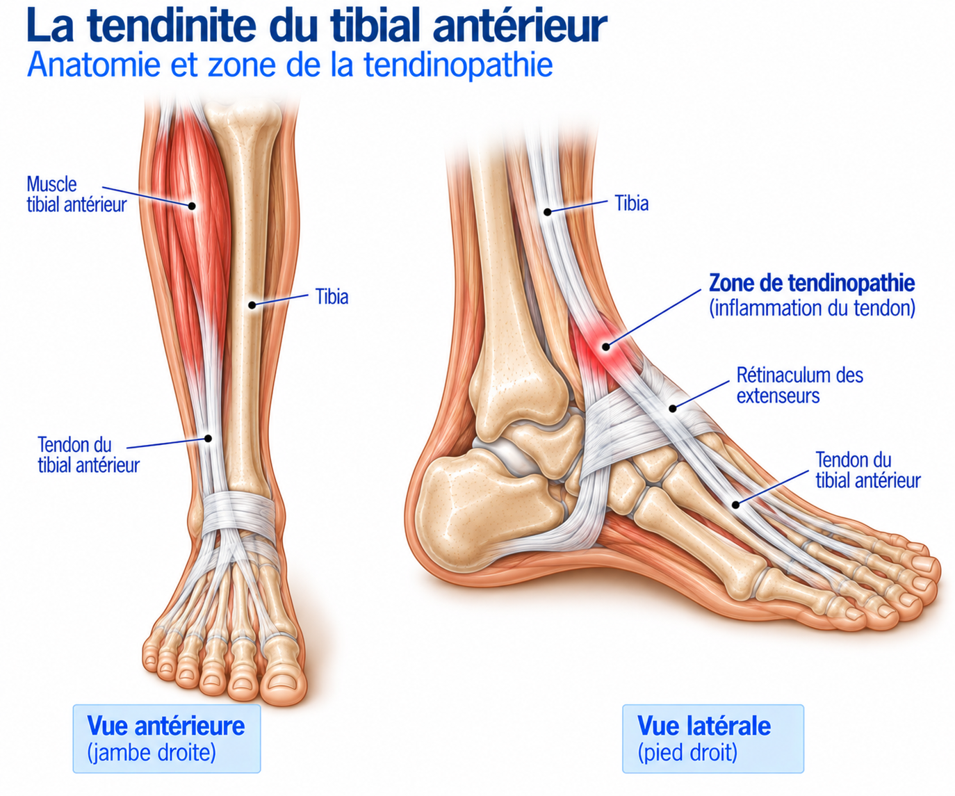
🔍 Click on the image to enlarge it
Function of the tibialis anterior
The tibialis anterior plays several essential roles: it produces dorsiflexion, that is, lifting the forefoot towards the leg, and contributes to inversion of the foot. During walking and running, it notably controls the lowering of the foot after initial contact, thereby contributing to load absorption, ankle stability, the forward progression of the tibia over the foot and preparing the foot for ground contact.
Biomechanics
The tibialis anterior works particularly hard during dorsiflexion, control of foot placement, brisk walking, running, uphill sections, some downhill sections and changes of pace. On ground contact, it contracts to gradually control the position of the foot: it therefore often works in eccentric contraction, that is, producing force while lengthening. This characteristic explains why an abrupt increase in running load can exceed the tendon’s capacity to adapt.
Epidemiology
Tibialis anterior tendinopathy is relatively rare, particularly compared with Achilles or tibialis posterior tendinopathy. It can nevertheless be seen in runners, trail runners, hikers, athletes whose activities involve a lot of elevation gain, and people returning to a demanding level of physical activity. It can also appear in less active patients after an unusual increase in their activity.
The available literature on this condition consists mainly of case series, narrative reviews and small studies: caution is therefore needed regarding the precise risk factors and the optimal treatment protocols.
Causes & risk factors
Increased load
The contributing factors are essentially mechanical. An abrupt increase in mileage or in the number of sessions, a return after time off, an increase in elevation gain, or the introduction of interval training or hill sessions can exceed the tendon’s capacity to adapt.
Change of surface
A change of surface — road to trail, flat to mountain, treadmill to outdoors, soft to hard ground — can alter the stresses placed on the tibialis anterior.
Change of shoes
A significant change in drop, stiffness, weight or geometry can alter the load on the ankle-foot complex. This does not mean that a particular shoe is responsible for the tendinopathy, but that an abrupt change can represent an additional increase in load.
The typical scenario in runners
A common scenario is a runner who abruptly changes their training — for example, a return to running combined with a rapid increase in mileage and several hill sessions. Pain then appears gradually: discomfort after running, then pain during running, then pain when walking or on stairs. This pain is often very localised to the front of the ankle.
Symptoms
The main symptom is pain on the front of the ankle, described as aching, pulling, burning, tightness, discomfort when flexing the foot or a feeling that the tendon is “irritated”. It can be provoked by running, brisk walking, uphill walking, climbing stairs, active dorsiflexion or lifting the foot against resistance.
The pain can be found in several places: most often over the distal portion of the tendon (front of the ankle, course of the tendon, the area just in front of the joint), sometimes at the insertion zone (medial cuneiform, base of the first metatarsal, medial midfoot), and more rarely at the tendon sheath, whose irritation can cause more diffuse pain with swelling, crepitus or a rubbing sensation.
A particularly suggestive finding is pain reproduced when the patient actively lifts the foot against resistance (“Pull your foot up towards you while I resist”), a manoeuvre that should always be compared with the opposite side. Stretching the tibialis anterior — with the foot gradually placed into plantar flexion with an eversion component — can also reproduce the usual pain, which is a further argument in favour of tendon involvement; the clinical literature also describes this passive stretch test as part of the examination.
Clinical examination
On inspection, the examiner looks for swelling, thickening of the tendon, asymmetry or a possible depression along the course of the tendon. Palpation looks for pain over the tendon, its sheath or its distal insertion. In contraction, active dorsiflexion, resisted dorsiflexion and resisted inversion are tested. On stretching, the examiner looks for reproduction of the usual pain.
The tibialis anterior should also be assessed in functional situations: walking, brisk walking, climbing stairs, running, walking uphill, walking downhill and dorsiflexion exercises. In runners, movement analysis can help identify the situations that increase the load on the tendon.
Warning sign not to be overlooked
Reduced dorsiflexion, marked weakness, a change in gait or “foot slap” (the forefoot dropping abruptly after ground contact, as if “the foot were falling” or “slapping” on the ground) should prompt investigation for a rupture of the tibialis anterior tendon rather than a conclusion of simple tendinopathy (see the Surgery section).
Further tests
Ultrasound
It allows assessment of tendon thickness, architecture and vascularity, and of any tenosynovitis, fissuring or rupture. It also allows a dynamic examination.
MRI
Can show tendon thickening, signal abnormalities, oedema, fissuring, involvement of the insertion or a rupture. A review specifically on tibialis anterior tendinopathy considers it an important diagnostic test when the diagnosis is uncertain or when a structural lesion is suspected.
Differential diagnoses
Extensor tendinopathy (big toe, toes)
Can cause pain in a nearby area, located over the adjacent extensor tendons rather than along the course of the tibialis anterior itself.
Extensor tenosynovitis
Can appear after an increase in running, overly tight shoes or unusual activity.
Anterior ankle impingement
Pain is provoked mainly by dorsiflexion, particularly forced dorsiflexion — of joint rather than tendon origin.
Ankle arthropathy
To be considered in cases of stiffness, swelling, deep pain or restricted joint movement.
Osteochondral lesion of the talus
Can cause deeper pain and sometimes mechanical symptoms (locking, catching).
Stress fracture
To be looked for in cases of very localised, gradually increasing bone pain, or pain at night or at rest.
Anterior compartment syndrome
To be considered when pain consistently appears during exercise, with a feeling of tightness in the compartment and possibly neurological symptoms.
Nerve entrapment (deep peroneal nerve)
Can mimic anterior ankle pain of neurological rather than tendon origin.
Rupture of the tibialis anterior tendon
Diagnosis is sometimes delayed because the toe and big toe extensors partially compensate for dorsiflexion. To be considered when there is reduced dorsiflexion, weakness, "foot slap", a change in gait or a mass/depression along the course of the tendon.
Treatment
General principle and acute phase: reducing irritability
Management follows the general principles for tendinopathies: temporarily reduce overload, control symptoms, gradually reload the tendon, restore strength, then gradually return to sport. As the literature specific to the tibialis anterior is limited, protocols are less standardised than for the Achilles tendon.
When pain is significant, running, elevation gain, speed, interval training and prolonged walking are temporarily reduced — the aim not necessarily being complete rest, but keeping up activities that do not make things worse. In very painful forms, temporary immobilisation may be offered (a stiffer shoe, a walking boot in some situations, a substantial reduction in stresses); reviews of foot and ankle tendinopathies cite it as an initial option in symptomatic forms of tibialis anterior tendinopathy, followed by progressive rehabilitation. However, this strategy must remain temporary: prolonged immobilisation leads to loss of strength, stiffness, reduced tendon capacity and deconditioning.
Gradual reactivation
As the pain decreases, the tendon is gradually reloaded: active dorsiflexion without resistance (3 × 15-20 repetitions), then dorsiflexion with light resistance (a light resistance band, gradually stronger), and isometric work holding dorsiflexion against resistance (for example 4-5 × 30-45 seconds). The aim is to gradually reintroduce a load the tendon can tolerate.
Progressive strengthening and eccentric / heavy slow resistance work
The load is then gradually increased: dorsiflexion with a resistance band, with a pulley, with weights, unilateral work and functional exercises — progression being guided by the tendon's response. When tolerance is sufficient, more demanding work can be introduced: slow dorsiflexion against resistance, slow return into plantar flexion, heavy slow resistance work, and control of the lowering of the foot. The aim is to restore strength, tendon capacity, endurance and neuromuscular control.
It should be stressed, however, that the data specific to the tibialis anterior are insufficient to claim that any particular eccentric protocol is superior to the others: the recommendations are largely extrapolated from the general principles of tendinopathy management.
Functional work for the foot and ankle
Brisk walking, climbing stairs, walking uphill, single-leg work, balance exercises and jumps are gradually reintroduced, followed by running, accelerations and changes of pace. As the tibialis anterior does not work in isolation, it is also useful to work on the intrinsic foot muscles, arch control, the triceps surae, the peroneal muscles and single-leg balance, as well as ankle mobility (dorsiflexion, plantar flexion, subtalar mobility), strength and proprioception. An ankle that can produce and absorb forces efficiently helps reduce excessive concentration of load on a single tendon.
Load management and gradual return to running
In runners, it is important to identify precisely what triggers the pain (speed, duration, elevation gain, surface, shoes, frequency of sessions). Cycling, swimming or the elliptical trainer can be continued during the recovery phase. The return follows a progression such as: walking → brisk walking → alternating walking/running → easy running → increased duration → increased volume → speed → hills → intervals, increasing only one variable at a time (for example, do not return simultaneously to your previous mileage, hills and intervals).
In a mild form, suitably adapted activity can sometimes be maintained by monitoring pain during the activity, how it changes over the session, and pain in the following hours and the next day. A gradual increase in pain or a loss of strength should lead to a reduction in load; significant pain combined with weak dorsiflexion or a suspected rupture, on the other hand, means running should be stopped and a medical assessment sought.
Complementary treatments
Shoes
There is no universally recommended shoe. During the painful phase, it may be preferable to choose a stable shoe, a sufficiently stiff sole, enough volume over the instep and no excessive compression of the tendon — a shoe that is too tight over the instep can increase local stresses on the extensor tendons and the tibialis anterior.
Insoles and orthoses
They are not routinely the first-line treatment. They may be considered when there is a clearly identified biomechanical abnormality that correlates with the symptoms, but should remain a complement: the main treatment remains load management, muscle reconditioning and functional progression.
Manual therapy
Can be used to improve a joint restriction, temporarily reduce pain, ease movement and support the return to exercise — but must not replace progressive loading of the tendon.
Shockwave therapy
Data specific to tibialis anterior tendinopathy are very limited, without sufficiently robust evidence to make it a standard treatment. It may possibly be considered in refractory chronic forms, with a level of evidence markedly lower than that available for other tendinopathies.
Corticosteroid injection
An injection directly into a tendon with structural damage is generally not favoured, because of the potential risk of weakening the tendon. An injection into the sheath may be considered in certain selected cases of tenosynovitis, but should not be regarded as a routine treatment.
PRP, prolotherapy, hyaluronic acid and other biological injections
The data specific to the tibialis anterior are insufficient to recommend them routinely. Isolated case reports exist (for example, percutaneous ultrasonic tenotomy reported after failure of other treatments in a resistant distal form with fissuring), but they do not allow a standard treatment strategy to be established.
Surgery
Surgery is rarely needed for simple tibialis anterior tendinopathy. It may be considered when pain persists despite well-conducted conservative management, when significant structural damage is identified, in the case of a diagnosed major partial rupture or complete rupture, or when the functional deficit is significant. A small surgical series of resistant tendinopathies showed improvement after debridement and tendon augmentation, but the data remain very limited.
Tibialis anterior rupture: a different situation
A complete rupture should be regarded as a condition distinct from tendinopathy. It can cause loss of dorsiflexion strength, “foot slap”, difficulty lifting the forefoot and a change in gait, with partial compensation by the toe and big toe extensors. The rupture may be traumatic (direct blow, wound, forced movement) or degenerative, occurring without major trauma in a previously weakened tendon — which is notably described in older patients. It is sometimes diagnosed late, precisely because function is partially compensated by the other extensor muscles.
Surgical treatment of rupture
In young, active patients with a complete rupture and functional deficit, surgical repair or reconstruction is frequently considered. The options depend on how long ago the rupture occurred, tendon quality, retraction and the patient’s activity. Techniques include direct repair, tendon transfer, autograft and reconstruction. A 2023 meta-analysis of 217 cases suggests better outcomes when ruptures are repaired in the acute phase, with favourable results for some direct repair or autograft techniques.
However, a rupture does not always require surgery: the decision depends in particular on age, activity level, functional deficit, how long ago the rupture occurred and tendon quality. In athletes with high functional demands, surgery is more readily considered.
Rehabilitation after surgery
Recovery depends heavily on the technique used and generally involves several stages: a protection phase (protecting the repair, immobilisation, oedema control, gradual return to weight-bearing according to the surgeon’s instructions), a phase of gradual recovery of ankle mobility, a progressive strengthening phase (tibialis anterior, triceps surae, peroneals, intrinsic foot muscles), a functional phase (balance, walking, climbing stairs, single-leg exercises) and then an impact phase (jumps, running, accelerations, sport-specific work).
Recovery time
The exact timeframe depends on the severity of the injury, the treatment given, any surgery, strength recovery and the sport practised: there is no single timetable that applies to all patients. A mild form may improve within a few weeks, whereas chronic forms or those with structural damage may require several months.
Before considering a return to running, the following are looked for: little or no pain, stable and not worse the next day; satisfactory dorsiflexion and functional mobility close to the opposite side; sufficiently strong dorsiflexion, good endurance and no significant functional deficit; and the ability to perform brisk walking, stair climbing, single-leg balance, jumps and dynamic exercises.
The progression for returning to sport can then follow: walking → jogging → continuous running → increased volume → accelerations → hills → intervals → sport-specific work.
Prevention
Prevention relies above all on sensible management of training load.
Avoid: an abrupt increase in mileage, an immediate return to your previous level, adding hills and intervals at the same time, and a radical change of shoes and surface at the same time.
Favour: gradual progression, regular strengthening, foot and ankle work, sufficient recovery and varied intensities.
Frequently asked questions
Is it tendinitis?
Is it common in runners?
Can I keep running?
How long does it take to heal?
Is an MRI essential?
Can a rupture go unnoticed?
Does a rupture always require surgery?
Common mistakes
Continuing to run despite gradually worsening pain: the tendon needs appropriate load, not repeated overload.
Immobilising for several weeks without rehabilitation: immobilisation can ease the pain but also reduces muscle capacity.
Treating with massage alone: it can provide relief but does not rebuild the tendon's capacity.
Doing stretching only: it does not replace strengthening.
Injecting directly into the tendon: the benefit/risk ratio is not favourable in simple tendinopathy.
Overlooking weak dorsiflexion: it may signal a much more serious injury, particularly a rupture.
Relying on pain alone: strength and function must also be assessed.
Key points — Institut Basque du Coureur
Tibialis anterior tendinopathy is a relatively rare condition, but one to be aware of in runners with anterior ankle pain, particularly when it is reproduced by resisted dorsiflexion. Management relies mainly on a temporary reduction in overload, symptom control, progressive strengthening, functional work and then a gradual return to running. Immobilisation can be useful in very irritable forms but must remain temporary. Imaging, particularly ultrasound or MRI, is especially valuable when the diagnosis is uncertain or when a structural lesion is suspected. Finally, weak dorsiflexion, foot slap or a clear change in gait should prompt investigation for a rupture of the tibialis anterior tendon rather than a conclusion of simple tendinopathy. The right treatment is not to protect the tendon indefinitely: it is to gradually rebuild its capacity to withstand the stresses the athlete wants to place on it.
- Harkin E, Pinzur M, Schiff A. Treatment of Acute and Chronic Tibialis Anterior Tendon Rupture and Tendinopathy. Foot Ankle Clin. 2017;22(4):819-831.
- Nery C, Alloza J, Bittar CK, et al. Tendinopathy of the tibialis anterior tendon: surgical management. Orthopade. 2010.
- Dean BJF, Getgood A, et al. Tendinopathies of the Foot and Ankle. Foot Ankle Clin. 2022.
- Vosoughi AR, Heyes G, Molloy AP, Mason LW, Hoveidaei AH. Management of tibialis anterior tendon rupture: Recommendations based on the literature review. Foot Ankle Surg. 2020;26(5):487-493.
- Christman-Skieller C, Merz MK, Tansey JP. A systematic review of tibialis anterior tendon rupture treatments and outcomes. Am J Orthop. 2015;44(4):E94-E99.
- Nery C, et al. Management of isolated anterior tibial tendon rupture: a systematic review and meta-analysis. Foot Ankle Int. 2019.
- Chen J, Kadakia R, Akoh CC, Schweitzer KM Jr. Management of Anterior Tibialis Tendon Ruptures. J Am Acad Orthop Surg. 2021;29(16):691-701.
- Tibialis Anterior Tendon Rupture Surgical Treatments and Outcomes: A Systematic Review and Meta-Analysis. J Foot Ankle Surg. 2023.
- Zielinska N, Tubbs RS, Paulsen F, et al. Anatomical Variations of the Tibialis Anterior Tendon Insertion: An Updated and Comprehensive Review. J Clin Med. 2021;10(16):3684.
- The tibialis anterior tendon revisited: a unified classification framework across development, evolution, and clinical application. Front Cell Dev Biol. 2025.
Physiotherapy protocol for this injury
Découvrez notre protocole de rééducation détaillé pour tibialis anterior tendinopathy : exercices adaptés, progression structurée et modalités de reprise.
Similar injuries
Tibial stress fracture
A tibial micro-fracture caused by repeated stress, requiring a mandatory break from running to allow the bone to heal.
Shin splints (medial tibial stress syndrome)
Inflammation of the membrane covering the tibia caused by repeated impacts, affecting 13 to 17% of runners.
Compartment syndrome
Increased muscle pressure during exercise that can compromise blood flow and nerve function.