
Definition
Shin splints, also known as Medial Tibial Stress Syndrome (MTSS), is a mechanical overload condition characterised by diffuse pain along the posteromedial border of the tibia. It occurs when the repeated stresses placed on the bone exceed its physiological capacity to adapt. This injury is particularly common in runners and is one of the main causes of lower leg pain in athletes.
Contrary to what its name suggests, shin splints is not simply an inflammation of the periosteum. Current scientific evidence shows that it is mainly a bone stress injury. When a runner increases training volume or intensity too quickly, bone remodelling becomes insufficient to repair the microstrain caused by repeated impacts: osteoclasts resorb bone faster than osteoblasts can rebuild it, leading first to bone microdamage and then to bone marrow oedema. The periosteum becomes painful secondarily, as a result of this bone distress and of the stresses exerted by the muscles that attach to the medial surface of the tibia.
Today, shin splints is considered to be part of the bone stress injury continuum, lying between the physiological adaptation of bone to training and tibial stress fracture. Without appropriate management, continued mechanical stress can lead to a true stress fracture. Early recognition of this condition is therefore essential to prevent this progression.
Anatomy
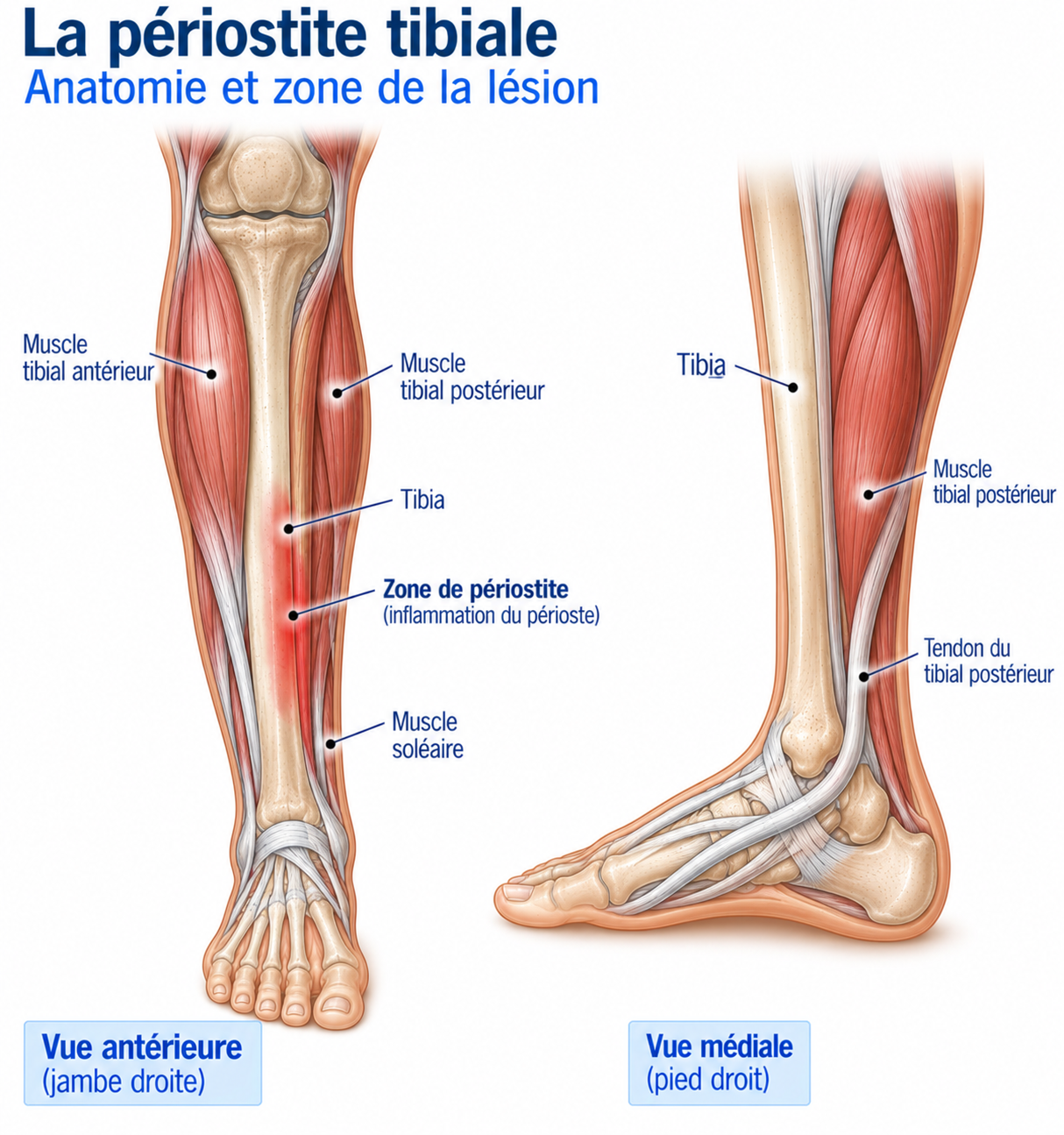
The tibia is the main weight-bearing bone of the lower leg and transmits loads between the knee and the foot during walking and running. With each stride, it withstands considerable forces generated by body weight, ground reaction forces and muscle contractions. The painful area in shin splints is most often located on the posteromedial surface of the tibia, in the middle or distal third of the bone, the area corresponding to the attachments of the soleus, tibialis posterior and flexor digitorum longus.
During running, these muscles exert repeated traction forces on the periosteum and help control ankle stability and support the arch of the foot. When stresses become excessive, the combination of bone compression forces and muscle traction promotes the onset of the pain characteristic of shin splints.
The soleus muscle originates from the posterior surface of the tibia and fibula before joining the Achilles tendon, enabling plantar flexion of the ankle and propulsion during running. The tibialis posterior muscle, for its part, arises from the posterior surface of the tibia, the fibula and the interosseous membrane before inserting mainly on the navicular and cuneiform bones: it is the main dynamic stabiliser of the medial arch of the foot and plays an essential role in controlling foot pronation. Finally, the flexor digitorum longus attaches to the posterior surface of the tibia and runs to the distal phalanges of the four lesser toes, contributing to propulsion and balance during the stance phase.
🔍 Click on the image to enlarge it
Biomechanics of the tibia during the running cycle
The tibia is subjected to considerable mechanical stress throughout the running cycle. In a runner, the forces transmitted to this bone can reach six to ten times body weight, depending on running speed, technique and surface. The bone acts as a true beam, able to absorb and then redistribute these stresses in order to limit their transmission to the joints.
At foot strike, a first stress peak occurs as ground reaction forces are transmitted to the skeleton; the lower leg muscles, notably the tibialis anterior and triceps surae, control this phase to limit the intensity of the impact. During the stance phase, the tibia bears almost all of the body weight and is simultaneously subjected to compression, bending and torsional stresses. During propulsion, the posterior lower leg muscles generate considerable force to move the body forward, which increases the traction stresses on the medial surface of the tibia.
In a healthy individual, these stresses stimulate bone remodelling and make the bone progressively stronger. When training volume or intensity increases faster than the bone tissue’s capacity to adapt, microdamage accumulates: the bone no longer has the time it needs to rebuild between sessions, which promotes the development of a bone stress injury.
Epidemiology
Shin splints is one of the most common overuse injuries in runners. Depending on the study, it accounts for 13 to 20% of running-related injuries, and this proportion can reach nearly 35% in military personnel undergoing intensive training. It affects beginner runners and experienced athletes alike.
This condition is seen in many sports involving repeated impacts (trail running, athletics, football, rugby, basketball, dance, gymnastics), and more often during marathon preparation, intensive training camps or a return to sport after a break.
Groups at higher risk include beginner runners, military personnel, women, and athletes with low bone mineral density, relative energy deficiency in sport (RED-S) or a history of stress fracture.
Causes & risk factors
Shin splints is a multifactorial condition: in the vast majority of cases, it does not result from a single factor but from a combination of several elements related to training, biomechanics, the runner’s morphology, their general health or their equipment. Identifying these factors is essential to prevent the injury and, above all, to avoid recurrence.
Training-related factors
The main risk factor is poor management of mechanical stress, resulting from an imbalance between volume, intensity and recovery. Increasing mileage too quickly, runs longer than usual, an abrupt return to sport or an accumulation of intense sessions do not give the bone enough time to adapt. Hills, long descents and interval training also increase the stresses placed on the tibia.
Insufficient rest between sessions, chronic fatigue or poor-quality sleep limit the capacity for bone remodelling.
Biomechanical factors
Weakness of the triceps surae, tibialis posterior, fibular muscles or gluteal muscles reduces the capacity to absorb stress. Limited ankle dorsiflexion and muscle stiffness alter the distribution of stress during running.
Impaired neuromuscular control, chronic ankle instability or poor pelvic stability promote increased internal rotation of the lower limb and increased foot pronation.
Stride characteristics
Prolonged overpronation increases the traction forces exerted by the tibialis posterior and the soleus. A low cadence combined with an excessively long stride (overstriding) increases ground contact time and the bending stresses on the tibia. Large vertical oscillation also increases impact energy.
Learn more
Changes to running gait can be an effective treatment when they are tailored to the runner’s profile and introduced gradually, ideally under the supervision of a professional. A moderate increase in cadence (5 to 10%) often reduces tibial stress without significantly changing running speed.
Static morphology
A flat foot with overpronation increases the stresses on the medial surface of the tibia, while a high-arched foot (pes cavus), which is more rigid, absorbs impacts less effectively. Calcaneal valgus or varus, excessive internal rotation of the tibia or a leg length discrepancy can also contribute to bone overload.
Individual factors
Low bone mineral density, vitamin D or calcium deficiency, relative energy deficiency in sport (RED-S), smoking, insufficient sleep, chronic stress or a pro-inflammatory diet reduce the capacity for bone repair. Some metabolic diseases, such as diabetes, can also delay remodelling.
Equipment factors
The choice of shoes is an important factor, although it is generally not solely responsible for the injury. Worn-out shoes, an abrupt change of model, too rapid a transition to minimalist shoes, insufficient cushioning or recently modified orthotic insoles can change the stresses placed on the tibia. Very hard or steeply sloping surfaces also increase mechanical stress.
Symptoms
Shin splints most often presents as gradually increasing pain along the posteromedial border of the tibia, in the middle or lower third of the leg. Symptoms develop progressively over several days or weeks, in connection with a recent increase in training load.
At first, pain appears only after running. Over time, it comes on earlier during exercise and may then persist throughout the session; in advanced forms, discomfort is present when walking, or even at rest.
The pain is usually diffuse, extending over more than five centimetres along the posteromedial border of the tibia, which helps distinguish it from a stress fracture, where pain is usually very localised. Patients frequently describe a burning, pulling or deep aching sensation, sometimes with morning stiffness.
Clinical examination
The clinical examination is above all a process of elimination, used to confirm the diagnosis and rule out a stress fracture or chronic compartment syndrome. Inspection rarely reveals any visible abnormality; slight oedema on the medial surface of the tibia may sometimes be observed, but it is inconsistent. Palpation reveals diffuse, reproducible pain along the posteromedial border of the tibia, over a length of more than 5 cm.
The biomechanical assessment systematically looks for contributing factors (overpronation, flat or high-arched foot, limited dorsiflexion, weakness of the triceps surae, tibialis posterior or gluteal muscles), as well as pelvic stability and running gait.
Two clinical tests are commonly used: the Hop Test (hopping on one leg, which often reproduces the pain) and the Single Leg Heel Rise Test (which assesses the strength and endurance of the triceps surae and tibialis posterior). Assessment of ankle mobility, particularly dorsiflexion, and checking for pain provoked by tapping the tibia or by weight-bearing usefully complete the examination. Neurological and vascular examination is normal in shin splints.
Further tests
Plain X-rays
Usually normal in the early stages. Mainly useful for ruling out another bone condition, but not very sensitive for detecting early stress injuries.
MRI
The reference investigation when the diagnosis is in doubt: it shows bone marrow oedema, periosteal oedema and the different stages of bone stress injury. Essential if a stress fracture is suspected.
Bone scintigraphy
Formerly widely used, now much less often prescribed because of the diagnostic superiority of MRI.
CT scan
Rarely indicated; useful in some complex stress fractures or when cortical analysis is needed.
Ultrasound
Limited role in diagnosing shin splints: mainly used to rule out an associated tendon or muscle condition.
Differential diagnoses
Tibial stress fracture
Very localised pain (often pinpoint, over a few mm to cm), which quickly becomes disabling and persists even during everyday activities, unlike the diffuse (> 5 cm) and progressive pain of shin splints.
Chronic exertional compartment syndrome
Pain appearing after a consistent amount of running time, with a marked feeling of muscle tightness, sometimes paraesthesia or muscle weakness, then rapid resolution after stopping exercise.
Other diagnoses to consider
Tibialis posterior tendinopathy, fibular tendinopathy, fibular stress fracture, tarsal tunnel syndrome, lumbar radiculopathy or, more rarely, a bone tumour or infection.
Classification
Stage 1
Pain appears only after sporting activity. Training can generally continue with a moderate reduction in load and prompt initiation of treatment.
Stage 2
Pain appears at the start of exercise, then eases during the warm-up before returning after the session. Training can be maintained if pain remains ≤ 3-5/10 and returns to its baseline level within 24h.
Stage 3
Pain is present throughout the activity and leads to reduced performance. Mechanical stresses must be reduced more substantially in order to prioritise rehabilitation.
Stage 4
Pain persists at rest or when walking. A stress fracture must always be looked for with an MRI; return to sport is suspended until a precise diagnosis has been made.
Treatment
Relative rest and load adjustment
Complete rest is rarely indicated: a well-reasoned adjustment of load generally allows faster recovery. In practice, running can continue if pain remains ≤ 3 to 5/10 during exercise, does not worsen as the session goes on and returns to its baseline level within 24 h. Otherwise, a greater reduction in stress is needed. Cycling, swimming, aqua jogging or the elliptical trainer help maintain fitness without impact.
Physiotherapy rehabilitation (first-line treatment)
Rehabilitation is the cornerstone of shin splints treatment. Its aim is not only to make the pain go away, but above all to gradually increase the capacity of the musculoskeletal system to withstand the stresses of running; it must be individualised according to the athlete's level, the risk factors identified during the initial assessment and the clinical course.
The first step is to reduce mechanical stress enough to allow bone remodelling to resume, without stopping all physical activity: keeping up non-impact activities (cycling, swimming, aqua jogging) preserves cardiovascular fitness while limiting the load on the tibia.
When pain allows, progressive strengthening is introduced following the principles of Heavy Slow Resistance (HSR) (slow movements under gradually increasing loads), targeting the triceps surae, tibialis posterior, fibular muscles, intrinsic foot muscles and gluteal muscles. Eccentric exercises are then added to the programme: they improve muscle control during the braking phases of the stride and tissue tolerance to repeated stress, but remain a complement to overall strengthening rather than the main treatment, unlike in tendinopathies.
Proprioception, motor control and pelvic stability play an important role in rehabilitation. Once muscle strength, mobility and motor control are satisfactory again, plyometric work (jumps, bounds, reactivity) is gradually introduced to re-accustom the bone to impact, an essential step before any full return to running. Finally, biomechanical correction (a moderate increase in cadence, shorter stride length, pelvic stability, foot strengthening) is offered according to the needs identified, without seeking to change the running gait systematically.
Gradual return to running
The return to running should never be guided solely by the disappearance of pain, but by the tissues' ability to gradually tolerate impact. Before any return, several criteria must be met: pain-free walking, strengthening performed without significant pain, Hop Test and hopping on the spot well tolerated, pain ≤ 3-5/10 during dynamic exercises.
The return begins with alternating walking and running (e.g. 1 min of running / 2 min of walking over about twenty minutes): if the session is well tolerated and pain returns to its baseline level in less than 24 h, running time is gradually increased over the weeks. The weekly volume increase should not exceed 5 to 10%. Intervals, hills and long runs are only reintroduced once continuous easy runs are completely pain-free.
Returning to the previous level of performance is often gradual and takes several weeks: returning too quickly carries a high risk of recurrence or progression to a stress fracture.
Complementary treatments
Orthotic insoles
May be offered to some runners with marked overpronation, flat feet or static morphological abnormalities, after clinical and biomechanical assessment. Not routine.
Extracorporeal shockwave therapy
May be considered in some chronic forms that do not respond to properly conducted management. Moderate benefit; the level of evidence is lower than for some tendinopathies.
Taping / strapping
Can be used temporarily to reduce certain biomechanical stresses or to reassure the patient during the return to activity. Its effect is mainly symptomatic.
Walking boot
Rarely necessary. May be considered in very painful forms or when a stress fracture is strongly suspected, while awaiting further investigations.
Non-steroidal anti-inflammatory drugs (NSAIDs)
Generally not recommended: as shin splints is a bone stress injury, inhibiting the inflammatory response could theoretically delay bone remodelling.
Corticosteroid injections
No indication in the management of shin splints; contraindicated because of the risk of weakening the tissues.
PRP (platelet-rich plasma)
To date, no high-quality study supports recommending PRP for this indication. Experimental use, outside current guidelines.
General factors in bone healing
Sufficient sleep, a balanced diet, adequate energy availability, correction of any vitamin D or calcium deficiency. Screening for RED-S should be considered in at-risk athletes (menstrual cycle disorders, significant weight loss).
Surgery
Surgery has virtually no role in the treatment of shin splints. The very rare indications involve persistent pain despite several months of perfectly conducted conservative treatment, and after all differential diagnoses have been ruled out. As results are variable, it is considered only as a last resort.
Recovery time
Recovery time depends on how early the diagnosis is made, the extent of the bone injury and adherence to rehabilitation principles. Early forms generally resolve well within 4 to 8 weeks when loads are adjusted promptly. More advanced forms often require 8 to 12 weeks, or even longer if the return is delayed. If there is a true stress fracture, recovery can take several months.
Return to sport should never depend solely on the time elapsed, but on meeting satisfactory functional criteria.
Prevention
Prevention relies above all on good management of training load: gradually increasing volume, intensity and frequency allows bone tissue to develop its capacity to adapt, while including sufficient recovery periods.
Regular strengthening of the triceps surae, foot muscles and glutes improves the capacity to absorb stress. Maintaining good ankle mobility, an appropriate running technique and a sufficient cadence also contribute to prevention.
Particular attention should be paid to sleep quality, nutrition and energy availability, as well as to screening for possible vitamin D deficiency or RED-S in at-risk athletes.
For a detailed approach, see our page “Preventing running injuries”.
Frequently asked questions
Can I keep running with shin splints?
How long do shin splints last?
Can shin splints progress to a stress fracture?
Do I need an MRI?
Are insoles always necessary?
Are anti-inflammatories useful?
Can I cycle?
Is shockwave therapy effective?
Does vitamin D play a role?
How can I prevent a recurrence?
Common mistakes
Continuing to train despite pain that is gradually increasing.
Stopping all activity completely for several weeks.
Immediately returning at the same level as before the injury.
Abruptly changing your shoes or running technique.
Neglecting strengthening.
Seeking only passive treatment without correcting the factors responsible for mechanical overload.
Key points — Institut Basque du Coureur
Shin splints is a bone stress injury resulting from an imbalance between the mechanical stresses placed on the tibia and its capacity to adapt. Early management based on load adjustment, progressive strengthening, correction of biomechanical factors and a well-reasoned return to running leads to full recovery in the vast majority of cases and limits the risk of progression to a stress fracture.
- Winters M, Moen MH, Zimmermann WO, et al. Medial Tibial Stress Syndrome: A Critical Review. Sports Med. 2018.
- Reinking MF. Exercise-related leg pain in female collegiate athletes. J Athl Train. 2006.
- Beck BR. Tibial stress injuries: an aetiological review. Sports Med. 1998.
- Brukner P, Khan K. Brukner & Khan's Clinical Sports Medicine. 6th ed. McGraw-Hill.
- Cook JL, Purdam CR. Is tendon pathology a continuum? Br J Sports Med. 2009.
- Malliaras P, Barton CJ, Reeves ND, Langberg H. Loading programmes in tendinopathy. Sports Med. 2013.
- JOSPT Clinical Practice Guidelines — Bone Stress Injuries. J Orthop Sports Phys Ther. 2024.
- ESSKA Consensus on Bone Stress Injuries in Athletes. 2023.
- Fredericson M, Jennings F, Beaulieu C, Matheson GO. Stress fractures in athletes. Top Magn Reson Imaging. 2006.
- Warden SJ, Davis IS, Fredericson M. Management and prevention of bone stress injuries. Br J Sports Med. 2021.
Physiotherapy protocol for this injury
Découvrez notre protocole de rééducation détaillé pour shin splints (medial tibial stress syndrome) : exercices adaptés, progression structurée et modalités de reprise.
Similar injuries
Tibialis anterior tendinopathy
Pain at the front of the ankle caused by the tendon of the muscle that lifts the foot.
Compartment syndrome
Increased muscle pressure during exercise that can compromise blood flow and nerve function.
Tibial stress fracture
A tibial micro-fracture caused by repeated stress, requiring a mandatory break from running to allow the bone to heal.