
Definition
Retrocalcaneal bursitis is a painful inflammation of the bursa located between the anterior surface of the Achilles tendon and the posterosuperior part of the calcaneus. The normal role of this small bursa is to help the Achilles tendon glide over the bone during ankle movements. In runners, it can become painful when it is subjected to repeated compression, shear and mechanical overload.
The term simply describes its location: retro (behind) + calcaneal (at the calcaneus) + bursitis (inflammation of a bursa). It lies deep to the Achilles tendon, which distinguishes it from superficial retro-Achilles bursitis, located between the tendon and the skin and much more directly influenced by friction or pressure from the shoe.
Retrocalcaneal bursitis is frequently associated with insertional Achilles tendinopathy, a posterosuperior prominence of the calcaneus (Haglund’s deformity), calcifications at the Achilles insertion, a sudden increase in running volume or intensity, or shoes that put significant pressure on the rearfoot. It is therefore important not to regard all pain at the Achilles tendon insertion as simple “tendinopathy”: the condition may mainly involve the bursa, the tendon, the bone, or several of these structures at the same time.
Anatomy
The back of the heel contains several closely interlinked structures: the Achilles tendon, the calcaneus, the retrocalcaneal bursa, the subcutaneous retro-Achilles bursa, the soleus muscle, the gastrocnemius, Kager’s fat pad, as well as the subtalar joint and surrounding structures.
The retrocalcaneal bursa lies between the anterior part of the Achilles tendon in front and the posterosuperior part of the calcaneus behind: it forms a true gliding interface between the tendon and the bone.
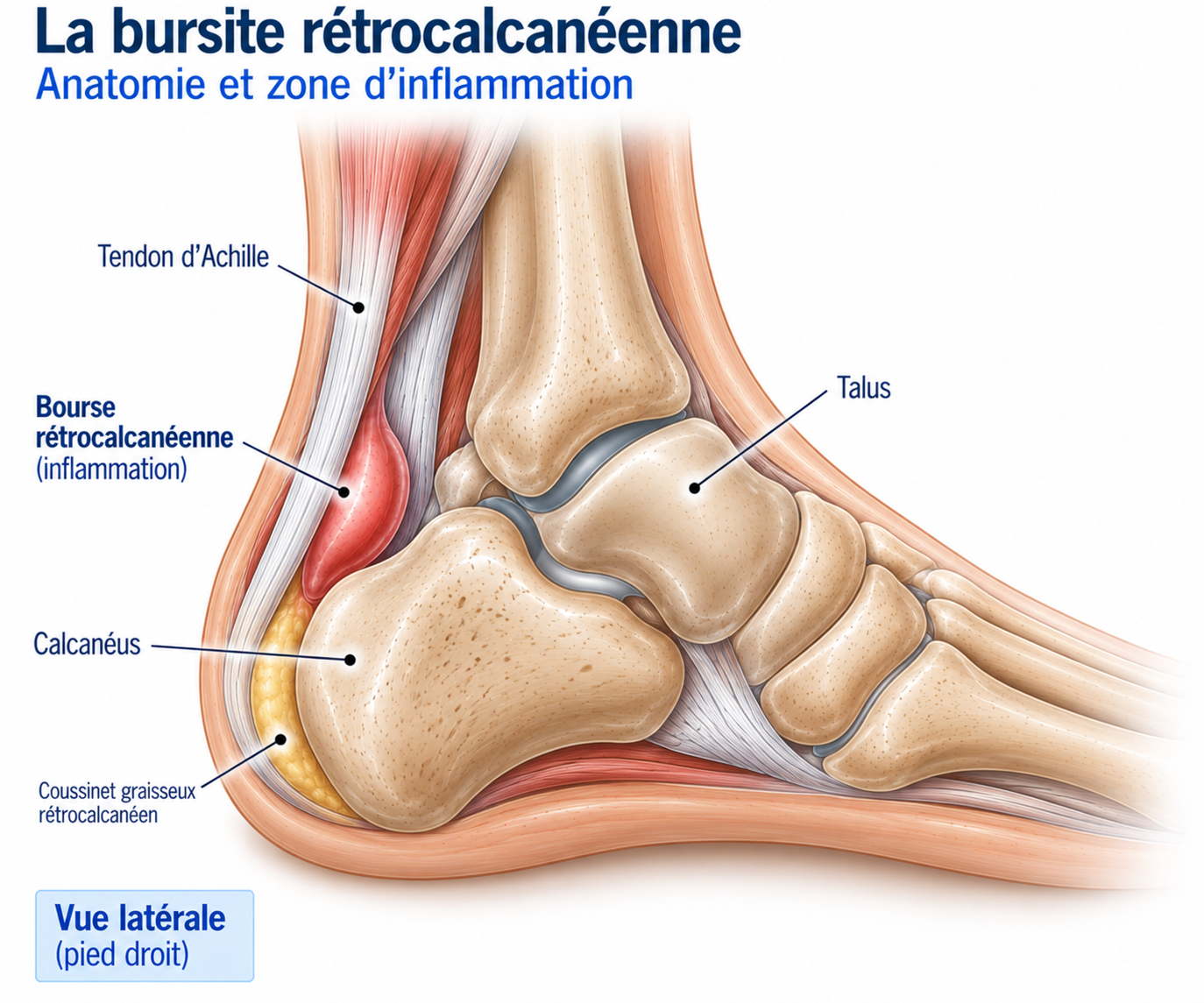
🔍 Click on the image to enlarge it
Role of the bursa and the vicious circle
The bursa normally allows the Achilles tendon to move over the calcaneus with minimal friction. When the ankle goes into dorsiflexion, particularly when the knee moves forward over the foot, the anterior part of the Achilles tendon and its bursa can be compressed more against the calcaneus. In an athlete with irritation in this area, this compression can become painful and sustain a vicious circle: compression → irritation → swelling of the bursa → less available space → further compression.
Biomechanics in runners
Running places considerable stress on the ankle-foot complex. With each stride, the foot makes contact with the ground, the ankle absorbs part of the load, the triceps surae gradually comes under tension, the Achilles tendon stores energy, and this energy is then released during propulsion. The tendon can withstand high loads; the problem arises when the tissues’ capacity to adapt is exceeded. Bursitis, however, is particularly influenced by local compression, and not only by the amount of force generated by the tendon.
Role of dorsiflexion
Dorsiflexion brings the Achilles tendon closer to the posterosuperior part of the calcaneus: marked dorsiflexion can therefore increase compression of the insertional region. This explains why some patients are particularly sore when going down stairs or a slope, during deep squats, or when stretching the calf and Achilles tendon. In bursitis or insertional tendinopathy, “stretching more” is therefore not necessarily the right strategy.
Epidemiology
Achilles pain is common in runners, but isolated retrocalcaneal bursitis is less common than Achilles tendinopathy: it most often occurs in the context of an associated insertional condition. It can be seen in distance runners, athletes in jumping sports, those who increase their load suddenly, or people returning to running after a break.
Some studies have found associations between calcaneal morphology, Achilles tendon thickness and the onset of bursitis in runners, but these anatomical factors should not be interpreted as sufficient causes on their own.
Causes & risk factors
Retrocalcaneal bursitis most often results from a combination of several factors related to training, biomechanics, footwear and the morphology of the rearfoot.
Training factors
Sudden increase in mileage or intensity, returning too quickly after a break, a large increase in hill work, more frequent interval or sprint sessions, more plyometric work, a sudden change of surface.
Biomechanical factors
Limited ankle mobility, increased dorsiflexion under load, a recent change in running technique, triceps surae weakness, a reduced capacity of the tendon to tolerate load.
Footwear-related factors
A shoe that is stiff or very high at the back can press directly on the Achilles region (external compression). Heel height and sole stiffness also change the stresses on the tendon: a slight heel raise can temporarily reduce dorsiflexion and therefore reduce insertional compression. A sudden change of shoes, an aggressive heel counter or minimalist shoes in a runner who is not prepared for them are additional risk factors.
Anatomical factors: Haglund’s deformity
Haglund’s deformity is a bony prominence of the posterosuperior part of the calcaneus. It can promote compression of the bursa, insertional tendinopathy and footwear-related pain. However, a bony prominence on imaging does not mean disease: some people have a Haglund morphology without any pain. Imaging must therefore always be correlated with the clinical findings — as with insertional calcifications or thickening of the Achilles tendon, which are also among the anatomical factors.
Symptoms
The main symptom is pain at the back of the heel, often deep, located just above the calcaneus, slightly lateral or medial to the tendon, and sometimes diffuse around the Achilles insertion. Patients typically say: “It hurts deep behind my heel” or “It hurts when I go downhill or when my ankle goes forward”.
Start-up pain may be present after a night’s sleep, a period of sitting, a car journey or a period of inactivity, and ease after a few minutes of walking. Unlike some tendinopathies, however, bursitis pain can remain very sensitive to mechanical compression.
When running, the pain may build gradually, appearing after several kilometres or from the very first minutes, and increase with intensity; accelerations and hills are often particularly provocative. Uphills place heavy demands on the triceps surae and can aggravate the pain; downhills require considerable eccentric control and greater ankle mobility, with dorsiflexion that increases retrocalcaneal compression. The classic calf stretch can also be particularly painful: in a symptomatic patient, aggressive stretching is therefore often best avoided at first.
Clinical examination
The examination looks for the exact location of the pain, tenderness on palpation, swelling, limited mobility, pain on dorsiflexion or on contraction of the triceps surae, involvement of the Achilles tendon, a calcaneal abnormality and footwear-related pain. Palpation must be precise: pain directly on the tendon at its insertion points more towards insertional tendinopathy, whereas deeper pain on either side of the tendon, particularly on compression of the retrocalcaneal region, points more towards bursal involvement — although the distinction is not always possible clinically.
The dorsiflexion test looks for reproduction of the pain by placing the ankle in dorsiflexion (knee straight, knee bent, weight-bearing or open chain), which is particularly suggestive in insertional conditions. The heel raise test (single-leg heel raise) assesses pain, strength, the ability to repeat the movement, asymmetry and movement quality; significant pain may indicate associated involvement of the Achilles tendon.
Further tests
Ultrasound
A particularly useful examination: it can show distension of the bursa, effusion, synovial thickening, hypervascularisation, associated insertional tendinopathy, calcifications or superficial bursitis. It also allows dynamic assessment of the region.
MRI
Useful when ultrasound is inconclusive, symptoms are atypical, deep or bone involvement is suspected, or surgery is being considered. It shows effusion and thickening of the bursal wall, inflammatory oedema, any insertional tendinopathy, bone oedema of the calcaneus, Haglund's deformity or calcifications.
X-rays
Useful for looking for a Haglund prominence, enthesophytes, calcifications, a bone abnormality or arthropathy — but they do not directly show inflammation of the bursa itself.
Differential diagnoses
Insertional Achilles tendinopathy
Very common and often associated with bursitis; pain arising mainly from the last few centimetres of the tendon and its insertion, provoked mostly by weight-bearing, uphills, accelerations, jumps and calf exercises.
Superficial retro-Achilles bursitis
More superficial, located between the tendon and the skin, and often directly related to footwear (friction, pressure).
Partial or complete Achilles tendon rupture
To be ruled out in the event of sudden pain, a snapping sensation, sudden loss of strength, difficulty rising onto the toes or an abnormal Thompson test.
Calcaneal stress fracture
Persistent pain, sometimes at night, or significant pain on direct compression of the calcaneus.
Posterior ankle impingement
Posterior pain triggered mainly by plantar flexion (and not by dorsiflexion).
Inflammatory disease (e.g. spondyloarthritis)
To be considered in cases of bilateral or recurrent retrocalcaneal bursitis, particularly if associated with inflammatory symptoms.
Classification
Stage 1 — Mild irritation
Mild pain, no pain at rest, activity still possible; pain mainly after heavy loads.
Stage 2 — Symptomatic bursitis
Pain during running, pain in dorsiflexion, discomfort with footwear, reduced performance.
Stage 3 — Significant bursitis / associated condition
Daily pain, significant functional limitation, large bursitis, associated insertional tendinopathy, significant bone abnormality.
Stage 4 — Resistant chronic form
Symptoms for several months, failure of well-conducted management, persistent mechanical abnormality, significant limitation in sport.
Treatment
Acute phase: reduce compression
Treatment rests mainly on three principles: reduce irritating compression, maintain musculotendinous capacity, then gradually rebuild load tolerance. The aim is not to put the tendon and bursa at complete rest, but to find a load low enough to allow recovery while maintaining the capacities of the musculotendinous complex.
During the first few weeks, the aim is to reduce forced dorsiflexion, calf stretching, steep hills, accelerations, jumps and exercises that strongly provoke pain. Running can be continued if it remains compatible with mild, stable pain. A comfortable shoe with a less aggressive heel counter, or even a small temporary heel lift to reduce dorsiflexion, can ease symptoms while strengthening work is resumed. Ice can be used as a symptomatic treatment after activity, and NSAIDs sometimes for a short period when acute pain is severe — without being the main treatment. Relative rest (removing the most irritating stresses while maintaining tolerated activities such as cycling or swimming) is sufficient: complete immobilisation is rarely necessary.
Progressive triceps surae rehabilitation (physiotherapy)
Physiotherapy is the core treatment: it makes it possible to control load, improve strength, restore mobility and gradually reintroduce sporting stresses. Progression starts with isometric contractions of the calf (a slight rise onto the toes, held for 20 to 45 seconds, 4 to 5 repetitions, avoiding excessive dorsiflexion), followed by concentric strengthening (double-leg then single-leg heel raises, with a gradual increase in repetitions, load and speed).
In retrocalcaneal bursitis, as in insertional tendinopathy, exercises in which the heel drops well below step level should be avoided at first, as this increases dorsiflexion and therefore compression: the work is initially done on flat ground or with a controlled range of motion. The soleus is then strengthened specifically (heel raises with the knee bent), before introducing heavy strengthening (weighted calf raises, calf press, single-leg work) and then single-leg work aiming for strength comparable to the unaffected side. Eccentric work can be used, but the rule remains to load the tendon without needlessly increasing its compression — recent data show that rehabilitation limiting tendon compression (limited dorsiflexion, avoiding aggressive stretching, a temporary heel lift) may be more effective, particularly when bursitis is present. The final stage, energy storage and release (small hops, jumps, skipping rope, progressive plyometric work), follows a progression from slow → fast, double-leg → single-leg, small range → large range, predictable → sport-specific.
A persistent increase in symptoms lasting more than 24 hours suggests that the load should be reduced: pain is monitored during activity, in the evening and the next morning, along with stiffness and the ability to perform the exercises.
Gradual return to running
The return to running can begin when walking is pain-free or almost pain-free, calf exercises are well tolerated, daily pain is under control and the patient can perform heel raises without significant pain. Start with slow running on flat ground. Hills are reintroduced once good musculotendinous capacity has been regained (gentle gradients, short duration, controlled speed, then progression). Intervals, accelerations and sprints are only reintroduced late, once good triceps surae strength and good tolerance of plyometric exercises have been regained.
Complementary treatments
Extracorporeal shockwave therapy
May be offered in chronic forms, particularly when insertional Achilles tendinopathy is also present. The evidence is stronger for tendinopathy than for isolated bursitis: they remain a complementary treatment, not a substitute for strengthening.
Ultrasound-guided injection of the bursa
May sometimes be considered when bursal inflammation is significant and persistent, with great caution. Injection directly into the Achilles tendon should be avoided, given the risk of weakening and rupture.
PRP (platelet-rich plasma)
May be offered for some Achilles tendinopathies, but the evidence is still insufficient for it to be a standard treatment for retrocalcaneal bursitis: it should not be offered routinely.
Surgery
Surgery is exceptional for isolated bursitis. It may be discussed when symptoms are chronic, functional impairment remains significant, several months of well-conducted conservative treatment have failed, a significant bone abnormality is sustaining the compression, or severe insertional tendinopathy is also present.
Procedures may include bursectomy, resection of the calcaneal prominence, treatment of Haglund’s deformity, tendon debridement, treatment of calcifications, or tendon repair/reattachment depending on the extent of the damage. An endoscopic approach may sometimes be considered for isolated bursitis, with potentially lower morbidity than open surgery. Surgery is not trivial, however (wound-healing problems, infection, persistent pain, nerve injury, adhesions, recurrence, tendon weakness or rupture) and should be reserved for properly selected patients — a Haglund abnormality on imaging is never, on its own, an indication for surgery.
Recovery time
Recent bursitis can resolve favourably within a few weeks thanks to reduced load, footwear adjustments, compression control and progressive rehabilitation. Chronic forms, particularly when associated with insertional tendinopathy or a bone abnormality, may require several months. The disappearance of pain and full recovery of sporting capacity do not always happen at the same time: the region must gradually regain its capacity to withstand compression, traction, high loads and energy storage/release, which takes time even after symptoms have disappeared.
Prevention
Prevention rests mainly on gradual training progression (avoiding sudden increases in mileage, elevation gain, speed or session frequency) and on regular strengthening of the gastrocnemius, soleus, foot muscles and posterior chain. When managing hills, it is important not to increase volume, speed and elevation gain at the same time, to avoid sudden changes of shoes, and to respect recovery periods between demanding sessions.
With age, the tendon’s capacity to adapt may decrease: for runners over 40, it becomes particularly worthwhile to keep 1 to 2 strengthening sessions a week, including heavy work, single-leg work and appropriate plyometric work. The best way to prepare a tendon to withstand the stresses of running is generally to expose it regularly and gradually to those stresses.
Frequently asked questions
Can I keep running with retrocalcaneal bursitis?
Is retrocalcaneal bursitis a form of tendinopathy?
Is cold therapy useful?
Is stretching recommended?
Can a heel lift help?
Does Haglund's deformity need surgery?
Can an injection be considered?
Is shockwave therapy effective?
How long does it take to heal?
Can you have bursitis on both sides?
Common mistakes
Stopping running completely for several months: rest alone does not restore the tendon's capacity.
Stretching the calf aggressively, which can increase compression of the insertional region.
Doing calf exercises on a step and letting the heel drop very low, which is particularly irritating at first.
Going straight back to hills, which place a heavy load on the triceps surae.
Changing shoes suddenly, which disrupts the load balance on the foot.
Treating only the inflammation, without restoring the mechanical capacity of the tendon and calf.
Injecting corticosteroids directly into the Achilles tendon.
Operating too quickly on Haglund's deformity when it is not necessarily the cause of the symptoms.
Key points — Institut Basque du Coureur
Retrocalcaneal bursitis is an irritation of the bursa located between the Achilles tendon and the calcaneus, often associated with insertional Achilles tendinopathy. The pain, deep and located at the back of the heel, is increased by dorsiflexion, hills, sprints and jumps, as well as by a shoe that compresses the back of the heel. Haglund's deformity can promote compression but is not always pathological. Treatment rests mainly on load management and progressive strengthening of the triceps surae, avoiding aggressive stretching and forced dorsiflexion at first; a heel lift can temporarily reduce compression. Intratendinous corticosteroid injections should be avoided, and surgery remains exceptional, reserved for resistant forms.
- Polzer H, Polzer S, Mutschler W, et al. Insertional Achilles tendinopathy: differentiated diagnostics and therapy. Orthopade. 2017;46:803-810.
- Wiegerinck JI, Kerkhoffs GMMJ, van Sterkenburg MN, Sierevelt IN, van Dijk CN. Treatment for insertional Achilles tendinopathy: a systematic review. Knee Surg Sports Traumatol Arthrosc. 2012;21:1345-1355.
- Van Dijk CN, van Dyk GE, Scholten PE, Kort NP. Endoscopic calcaneoplasty. Am J Sports Med. 2001;29:185-189.
- Chimenti RL, Cychosz CC, Hall MM, Phisitkul P. Current concepts review update: insertional Achilles tendinopathy. Foot Ankle Int. 2017;38:1166-1175.
- McAlister JE, Granata JD, Sennett BJ, et al. Management of insertional Achilles tendinopathy. J Am Acad Orthop Surg. 2022;30:e600-e609.
- Wiegerinck JI, Kok AD, van Dijk CN. Surgical treatment for insertional Achilles tendinopathy and retrocalcaneal bursitis. Foot Ankle Clin. 2014;19:741-755.
- Pringels L, Capelleman R, Van den Abeele A, et al. Effectiveness of reducing tendon compression in the rehabilitation of insertional Achilles tendinopathy: a randomised clinical trial. Br J Sports Med. 2025;59:640-650.
- Zhang C, Cao J, Yang L, Duan X. Surgical treatment for insertional Achilles tendinopathy and retrocalcaneal bursitis: more than 1 year of follow-up. J Int Med Res. 2021;49:300060521992959.
- Pierrart C, et al. MRI of the Achilles tendon: a comprehensive review of the anatomy, biomechanics, and imaging of overuse tendinopathies. Skeletal Radiol. 2010.
- Kose O, et al. Ultrasound-guided interventions in Achilles tendon and plantar fascia disorders. J Clin Med. 2023.
- Superficial retrocalcaneal bursae and nerves: from anatomy to ultrasound-guided procedures. Clin Anat. 2024.
- Surface shape of the calcaneal tuberosity and the occurrence of retrocalcaneal bursitis among runners. Medicina. 2021.
Similar injuries
Plantar plate injury
Damage to the fibrous plate that stabilises the toes, causing pain under the forefoot and a feeling of walking on a pebble.
Peroneal (fibularis) tendinopathy
Irritation of the lateral stabilising tendons of the ankle.
Plantar fasciopathy
Heel pain caused by irritation of the plantar fascia, very common in runners.