
Definition
Plantar fasciitis, also known as plantar fasciopathy, is one of the main causes of heel pain in adults. It refers to pain usually located at the proximal insertion of the plantar fascia on the calcaneus, the most common form being proximal plantar fasciopathy, located close to the medial tubercle of the calcaneus.
It affects both people who take part in sport and sedentary individuals. In athletes, it is particularly common in activities involving repeated impacts: running, trail running, team sports, tennis, dance or activities involving jumping.
The term “fasciitis” suggests pure inflammation. However, in chronic forms, the changes observed in the fascia correspond more closely to a fasciopathy related to mechanical overload, with collagen disorganisation, increased thickness and reduced mechanical capacity of the tissue. The problem can therefore be summarised as an imbalance between the capacity of the fascia to withstand stress and the mechanical stresses placed on it.
Anatomy
The plantar fascia is a thick fibrous structure located under the foot, extending from the calcaneus towards the toes. It has three main bands: a medial band, a central band — the thickest and most important biomechanically — and a lateral band. It attaches at the back to the medial part of the calcaneus and then gradually divides towards the toes.
It is closely related to the intrinsic foot muscles, the flexor tendons, the metatarsophalangeal joints, the plantar fat pad and the calcaneus. Its role is not limited to protecting the structures under the foot: it actively contributes to stability of the arch and force transmission during walking and running.
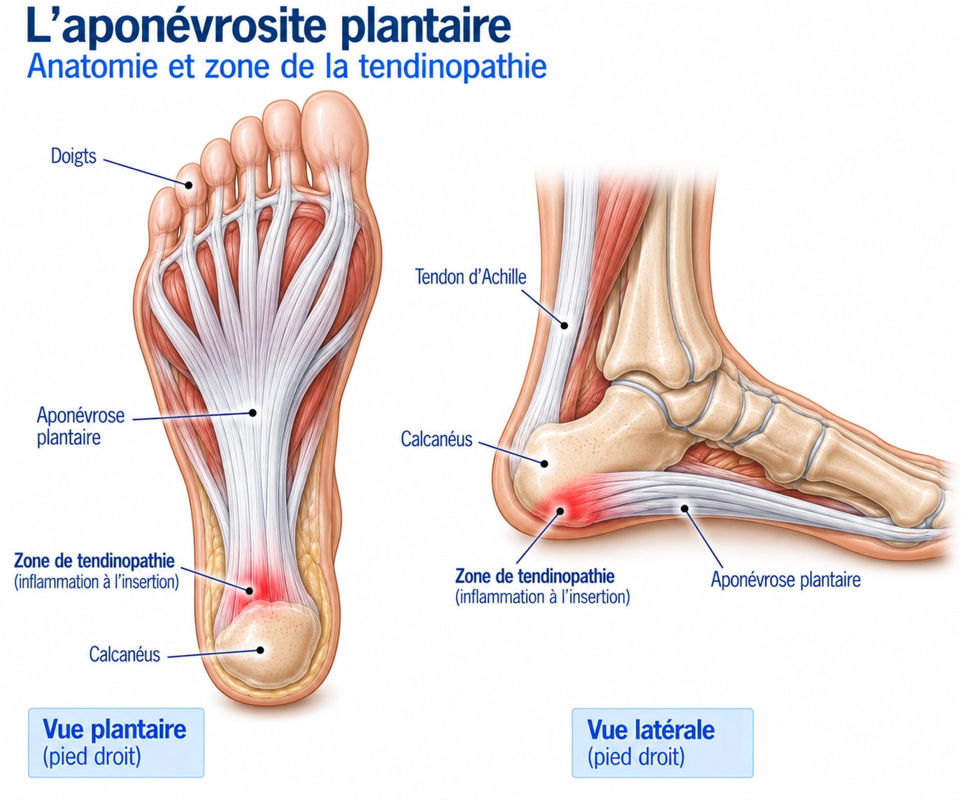
🔍 Click on the image to enlarge it
The Windlass mechanism
The plantar fascia works like an elastic system. During loading, it helps control the lowering of the arch; during propulsion, it helps stiffen the foot. This function is explained by the windlass mechanism: when the toes extend, particularly the big toe, the plantar fascia winds around the metatarsal heads. This tension raises the arch, shortens the functional length of the foot, stiffens the foot and promotes force transmission during propulsion. This mechanism is essential for running: when the plantar fascia is subjected to repeated stresses that exceed its capacity to adapt, microtears may appear and then accumulate.
The role of the triceps surae
The function of the plantar fascia is closely linked to that of the gastrocnemius + soleus + Achilles tendon + foot complex. Reduced ankle mobility, particularly dorsiflexion, can increase the stresses placed on the foot during stance and propulsion: marked stiffness of the triceps surae may therefore be a contributing factor in some patients. However, limited dorsiflexion should not be considered a single cause — the assessment must take the whole functional chain into account.
Epidemiology
Plantar fasciopathy is one of the most common causes of heel pain. It may account for up to around 10% of running-related injuries, depending on the populations studied. It mainly affects middle-aged adults but can also affect younger athletes.
Runners, people who are overweight and individuals exposed to long periods of standing are among the populations at risk.
Most patients make good progress with conservative treatment, but some forms become chronic and can last several months.
Causes & risk factors
Plantar fasciopathy is a multifactorial condition: it usually results from a combination of several factors related to training load, foot biomechanics and individual characteristics.
Load-related factors
The main triggering situations are a sudden increase in mileage, a return to running after a break, an increase in the number of sessions or in speed sessions, the introduction of hills, more jumping, a change of running surface or more time spent standing. In runners, a recent change to the training programme is very frequently found.
Biomechanical factors
Several characteristics can alter the stresses placed on the fascia: limited ankle dorsiflexion, reduced triceps surae strength, marked pronation, flat feet, high-arched (cavus) feet, reduced capacity of the intrinsic foot muscles or impaired foot control. However, it is important not to regard a particular morphology as pathological in itself: a flat foot or a high-arched foot can be completely asymptomatic. The deciding factor is often the capacity of the foot to withstand the stresses placed on it.
Individual factors
Several factors are associated with increased risk: age, being overweight, prolonged standing, a job involving a lot of walking, diabetes, reduced ankle mobility or a history of plantar fasciopathy. In athletes, low strength of the triceps surae and the intrinsic foot muscles can also contribute to overload.
Symptoms
The characteristic symptom is pain under the heel, usually on its medial side. Patients very often describe severe pain with the first steps in the morning, which may gradually ease after a few minutes of walking. It can also appear after sitting for a long time, with the first steps after a car journey, after a period of rest, while running or after prolonged standing — this feature is particularly suggestive.
At first, the pain mainly appears with the first steps and then gradually disappears. As the condition progresses, it may return during walking, appear while running, persist after exercise, or even be present at rest in severe forms. A particularly important element is the foot’s reaction the day after an activity: an increase in morning pain after a session is often an indicator that the load was too high.
Clinical examination
The diagnosis is mainly clinical. Palpation looks for pain at the medial tubercle of the calcaneus and the proximal insertion of the fascia; the pain is usually very localised. The examination also looks for pain on passive extension of the toes, limited ankle dorsiflexion, stiffness or weakness of the triceps surae, weakness of the intrinsic foot muscles, an abnormality of the arch or pain on weight-bearing.
The Windlass test involves tensioning the fascia by extending the toes, particularly the big toe: reproducing the pain at the calcaneal insertion can support the diagnosis, but no single test can confirm or rule out plantar fasciopathy on its own.
The functional assessment looks at the patient’s capacity to tolerate different loads: single-leg heel raise (triceps surae strength and endurance, foot control), single-leg balance (foot and ankle stability), single-leg squat (ankle mobility, control of the lower limb and pelvis) and, in athletes, jumps to assess the foot’s capacity to withstand rapid loading.
Further tests
Plain X-rays
May reveal a heel spur in some patients. However, this bony outgrowth is not necessarily responsible for the pain: it can be present in asymptomatic individuals and is not, on its own, an indication for surgery.
Ultrasound
May show thickening of the plantar fascia, reduced echogenicity, fibrillar disorganisation or hypervascularisation. A thickness greater than approximately 4 mm is often suggestive, but ultrasound abnormalities can exist in asymptomatic patients.
MRI
Allows examination of the plantar fascia, calcaneus, soft tissues, muscles, nerves and joint structures. Particularly useful when a stress fracture or another cause of heel pain is suspected.
Differential diagnoses
Calcaneal stress fracture
Pain often more diffuse on palpation around the calcaneus, in a context of marked training overload; to be considered in the presence of unusual pain at rest or at night.
Heel fat pad syndrome
Pain more centred under the heel, made worse by direct weight-bearing and hard surfaces, with no clear link to the first steps in the morning.
Tibial nerve neuropathy / tarsal tunnel syndrome / inferior calcaneal nerve
Pain that may be accompanied by paraesthesia, burning or electric-shock sensations, sometimes made worse by prolonged standing or at night.
Tibialis posterior or peroneal (fibularis) tendinopathy
Pain located more behind or below the malleolus, reproduced by stretching or resisted contraction of the tendon concerned rather than by palpation of the calcaneal tubercle.
Sever's disease (children)
Heel pain in children or adolescents during growth, related to the calcaneal apophysis rather than to the plantar fascia.
Inflammatory arthropathy / osteoarthritis / tumour or infection
To be considered in the presence of marked night pain, significant oedema, neurological signs, a history of trauma or an atypical course — should prompt a reassessment of the diagnosis.
Classification
Stage 1
Pain mainly present during the first steps. Everyday walking remains well tolerated.
Stage 2
Pain present during prolonged or sporting activities. Function remains largely preserved.
Stage 3
Pain present during walking and limiting sporting activities. A significant adaptation of load becomes necessary.
Stage 4
Severe pain in everyday life, sometimes present at rest. The diagnosis should be reassessed and differential diagnoses investigated.
Treatment
Load adjustment and pain monitoring
The first step is to identify the activities that exceed the foot's current capacity. It may be necessary to temporarily reduce running, jumping, long walks, hiking or prolonged standing. Complete rest is generally not necessary: activities without significant impact (cycling, swimming, upper body strengthening, suitable cardiovascular exercise) can be continued.
As with other overuse conditions, pain can be used as a load indicator. Moderate pain during exercise may be acceptable; the key element is the foot's reaction in the following hours and above all the next morning. A practical rule is to aim for pain that remains stable during activity, without a progressive increase, a return to the usual level within 24 hours and no worsening of first-step pain the next day. Morning pain is therefore an excellent practical marker of load tolerance.
Physiotherapy rehabilitation (core treatment)
Rehabilitation begins with gentle exercises aimed at reducing symptoms and restoring a minimum load capacity: isometric contractions, ankle mobilisation, light triceps surae strengthening and activation of the intrinsic foot muscles.
Strengthening of the triceps surae and the foot is then progressed: double-leg heel raises, then single-leg, then with added load, along with strengthening of the intrinsic muscles. A particularly well-studied protocol involves performing heel raises with the toes extended, in order to gradually increase the load applied to the fascia and restore its capacity to withstand the stresses of the Windlass mechanism; the progression follows the sequence bilateral → unilateral → added load → speed and power.
Gastrocnemius and soleus stretches may be offered to patients with limited ankle mobility, without causing significant pain at the fascia insertion. A specific plantar fascia stretch can also be performed by gradually extending the big toe while keeping the ankle in dorsiflexion, partly reproducing the Windlass mechanism — a particularly useful technique for patients with severe morning pain, to be done regularly and progressively.
Once strength is sufficient, rehabilitation moves on to dynamic function: single-leg balance, squats, lunges, step-ups, step-downs, walking on tiptoe. In athletes, double-leg and then single-leg jumps, bounds, skipping rope and plyometric exercises are then gradually introduced, in order to gradually re-accustom the fascia and the whole foot to rapid loading.
Gradual return to running
Before returning to running, walking should ideally be well tolerated, the first steps in the morning should be clearly improved, heel raises should be performed correctly, dynamic exercises should be tolerated and pain should remain stable after sessions.
The return can begin with an alternation such as 2 minutes walking / 1 minute running, with the total duration adapted to the athlete's level. Progression can then be considered when pain during exercise remains controlled, first-step pain does not worsen the next day, heel raises are well tolerated, strength is improving, dynamic exercises are performed correctly and prolonged walking becomes pain-free or almost pain-free. The next-morning symptom remains one of the best practical indicators of load tolerance throughout the return.
Complementary treatments
Foot orthoses
Can support the arch, change pressure distribution and make it easier to resume activity. Recent data suggest a favourable effect on pain in some patients but a more limited benefit on function: to be used alongside active management, never as the only solution.
Temporary footwear adaptation
Comfortable shoes, good foot support, a stable sole, appropriate cushioning and possibly a slight drop during painful phases. Abrupt changes of shoes, particularly to a minimalist model, should be avoided.
Taping
Some techniques designed to support the arch can reduce pain in the short term, particularly when resuming activity. The effect is mainly symptomatic, alongside exercises — it does not replace strengthening.
Extracorporeal shockwave therapy
Can be considered in persistent forms after several months of well-conducted conservative treatment. Results vary depending on the protocols and meta-analyses: to be considered a complementary treatment, not a replacement for rehabilitation.
Corticosteroid injections
Can provide rapid pain relief, but the benefit is mainly short-term. Repeated use should be avoided because of the risk of fat pad atrophy, weakening of the plantar fascia and rupture: they are not a long-term treatment.
PRP (platelet-rich plasma)
Offered in some resistant chronic forms, with mixed results due to highly variable protocols. It is not a first-line treatment; it may be discussed after failure of well-conducted conservative management.
Temporary immobilisation
May be discussed in some very painful forms where walking is very difficult, while another condition is being ruled out. It must remain temporary and be followed by gradual rehabilitation.
Surgery
Surgery is only exceptionally necessary. It may be discussed in very disabling chronic forms, after several months of properly conducted conservative treatment. Procedures may include debridement of the diseased plantar fascia, partial release, partial fasciotomy or treatment of an associated abnormality.
Excessive release of the plantar fascia should be avoided: it can alter foot biomechanics and lead to destabilisation of the arch. Surgery should therefore be reserved for resistant forms and performed after a full reassessment of the diagnosis.
Recovery time
Plantar fasciopathy is a condition that can take a particularly long time to recover from. Improvement may be seen within a few weeks, but symptoms can persist for several months. However, most patients make good progress with conservative treatment.
Recovery depends in particular on how long the symptoms have been present, the level of pain, body weight, sporting load, ankle mobility, triceps surae strength and adherence to rehabilitation. Rapid disappearance of pain does not necessarily mean that the foot has immediately regained its maximum load capacity.
Prevention
Prevention is based mainly on a gradual progression of training load, regular strengthening of the triceps surae and the intrinsic foot muscles, maintaining good ankle mobility, gradual adaptation to footwear and a gradual return after a break.
In runners, certain periods carry a particularly high risk: returning after an injury, marathon preparation, a rapid increase in mileage, taking up trail running, increasing elevation gain or switching to minimalist shoes. Greater vigilance during these periods helps limit the risk of onset or recurrence.
Frequently asked questions
Can I keep running with plantar fasciopathy?
Why does it hurt mostly in the morning?
Is it really an inflammation?
Should I use ice?
Are insoles essential?
Should I stretch the plantar fascia?
Is shockwave therapy effective?
Can an injection be considered?
Does the heel spur need to be removed?
How long does it take to heal?
Can recurrences be prevented?
Common mistakes
Stopping all activity completely for several weeks.
Resuming abruptly as soon as the pain disappears.
Relying on stretching alone without strengthening work.
Neglecting strengthening of the triceps surae and the intrinsic foot muscles.
Changing shoes abruptly.
Using orthopaedic insoles without accompanying rehabilitation.
Piling up passive treatments, in particular repeated corticosteroid injections.
Systematically assuming the heel spur is the cause of the pain.
Ignoring atypical or treatment-resistant pain without reassessing the diagnosis.
Key points — Institut Basque du Coureur
Plantar fasciitis, or plantar fasciopathy, is above all an overload condition of the fascia located under the foot. The characteristic pain of the first steps in the morning is highly suggestive of the diagnosis. Treatment is based mainly on temporary load adaptation, gradual strengthening of the triceps surae and the foot muscles, and specific stretches of the plantar fascia and the calf. Insoles, taping and shockwave therapy can be used as complementary treatments in some patients. Recovery is generally gradual and may take several weeks to several months.
- Koc TA Jr, Bise CG, Neville C, Carreira D, Martin RL, McDonough CM. Heel Pain—Plantar Fasciitis: Revision 2023. J Orthop Sports Phys Ther. 2023;53(12):CPG1-CPG39.
- Trojian T, Tucker AK. Plantar fasciitis. Am Fam Physician. 2019;99(12):744-750.
- Martin RL, Davenport TE, Reischl SF, et al. Heel Pain—Plantar Fasciitis: Revision 2014. J Orthop Sports Phys Ther. 2014;44(11):A1-A33.
- Thomas JL, Christensen JC, Kravitz SR, et al. The diagnosis and treatment of heel pain: a clinical practice guideline—revision 2010. J Foot Ankle Surg. 2010;49(3 Suppl):S1-S19.
- Simental-Mendía M, Simental-Mendía LE, Sánchez-García A, et al. Effect of extracorporeal shockwave therapy on plantar fascia thickness in plantar fasciitis: a systematic review and meta-analysis of randomized controlled trials. Arch Orthop Trauma Surg. 2024;144:3503-3516.
- Lippi L, Folli A, Moalli S, et al. Efficacy and tolerability of extracorporeal shock wave therapy in patients with plantar fasciopathy: a systematic review with meta-analysis and meta-regression. Eur J Phys Rehabil Med. 2024;60(5):832-846.
- Heide M, Røe C, Mørk M, et al. Radial extracorporeal shock wave therapy and exercise combined with advice and customised foot orthoses for plantar fasciopathy: a randomized controlled trial. Br J Sports Med. 2024;58:910-918.
- Cortés-Pérez I, Moreno-Montilla L, Ibáñez-Vera AJ, et al. Efficacy of extracorporeal shock waves compared with corticosteroid injections in plantar fasciitis: systematic review and meta-analysis. Clin Rehabil. 2024;38:1023-1043.
- Gerwin R, et al. Effects of therapeutic interventions on pain due to plantar fasciitis: a systematic review and meta-analysis. J Clin Med. 2023.
- Systematic review and meta-analysis of foot orthoses for pain and function in plantar fasciitis. 2025.
Physiotherapy protocol for this injury
Découvrez notre protocole de rééducation détaillé pour plantar fasciopathy : exercices adaptés, progression structurée et modalités de reprise.
Similar injuries
Sinus tarsi syndrome
Inflammation of the anatomical cavity between the talus and the calcaneus, often following a poorly rehabilitated ankle sprain.
Retrocalcaneal bursitis
Inflammation of the bursa between the Achilles tendon and the calcaneus, causing pain at the back of the heel in runners.
Blisters in runners
Fluid-filled pockets under the skin caused by friction, heat and moisture, common in running, trail running and marathons.