
Definition
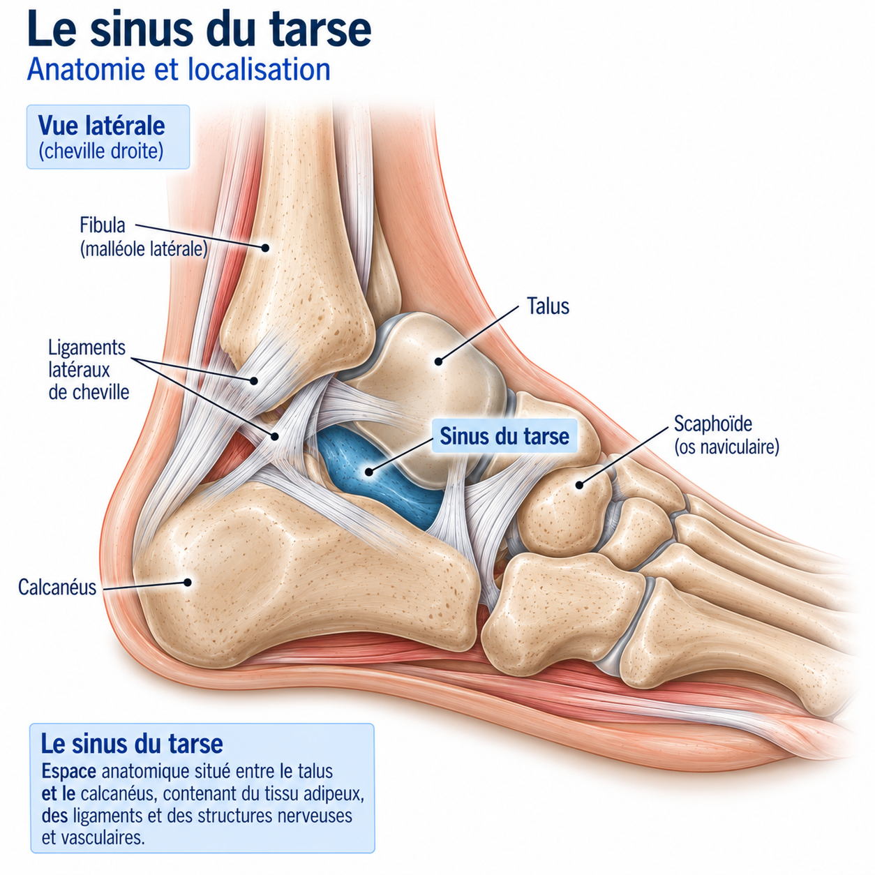
Sinus tarsi syndrome refers to pain located on the anterolateral part of the hindfoot, at the sinus tarsi, often associated with a feeling of hindfoot instability or of the “ankle giving way”. The sinus tarsi is an anatomical space located between the talus and the calcaneus, on the lateral side of the foot.
Historically, this syndrome was mainly considered to be the result of inflammation of the sinus tarsi after a sprain. Current evidence shows a more complex reality: the term may in fact cover several different situations — subtalar instability, injury to the talocalcaneal ligaments, injury to the cervical ligament, synovitis of the subtalar joint, fibrosis of the sinus tarsi, impingement, subtalar arthropathy, after-effects of an ankle sprain or morphological abnormalities of the foot.
It is therefore preferable, whenever possible, to identify the precise cause rather than to consider the syndrome as a single diagnosis.
Anatomy
Runners place enormous demands on the hindfoot. With each stride, the foot must in turn absorb stress, adapt to the terrain, control pronation, stiffen and then transmit forces towards the forefoot. The subtalar joint plays a major role in this adaptation; when it becomes painful or unstable, the runner may feel deep lateral pain, a sense of insecurity, episodes of “turning the foot”, difficulty running on uneven ground or discomfort on bends and cambered slopes.
The sinus tarsi is a tunnel-shaped space located between the talus and the calcaneus, on the lateral side of the hindfoot, just in front of the lateral malleolus. It contains fatty tissue, blood vessels, nerve endings and ligamentous structures, as well as some of the stabilising structures of the subtalar joint. It therefore plays an important role in proprioception and hindfoot stability.
🔍 Click on the image to enlarge it
The subtalar joint
The subtalar joint comprises the articulations between the talus and the calcaneus. In particular, it contributes to the combined movements of inversion, eversion, pronation and supination, which cannot be reduced to a simple rotation around an axis. It thus helps the foot adapt to uneven terrain.
The ligaments of the sinus tarsi
Several ligaments contribute to the stability of the region, including two particularly important structures: the cervical ligament, which contributes to the stability of the anterior part of the subtalar joint, and the interosseous talocalcaneal ligament, located deep in the tarsal canal, which plays a major role in stability between the talus and the calcaneus. An injury to these structures can lead to an abnormal increase in subtalar movement.
The role of proprioception
The sinus tarsi contains numerous mechanoreceptors that contribute to perceiving the position of the foot, hindfoot movements, changes in terrain and joint stresses. After a sprain, impaired proprioception may contribute to the feeling of instability: the problem is therefore not only mechanical, it may also be neuromuscular and proprioceptive.
Epidemiology
The exact frequency of sinus tarsi syndrome is difficult to determine. Terminology is not consistent across the literature, and the syndrome probably covers several different conditions.
It is, however, frequently described in athletes with recurrent sprains, chronic ankle instability, subtalar instability or persistent hindfoot pain. Surgical series show a strong association with a history of trauma, particularly inversion sprains.
Causes & risk factors
One of the main associations of sinus tarsi syndrome is subtalar instability, which may develop after a severe sprain, recurrent sprains, a ligament injury, an inversion injury or, more rarely, a non-traumatic condition. Instability can lead to excessive movement of the calcaneus relative to the talus, and these repeated movements can set off a genuine vicious circle: microtrauma → synovitis → fibrosis → pain → proprioceptive deficit → further instability.
The classic mechanism
The typical scenario is a runner who has had an inversion sprain, which may injure the lateral ankle ligaments, the cervical ligament, the interosseous talocalcaneal ligament, or several structures at once. The ankle may then seem to have “healed”, but the athlete continues to feel deep pain, instability, discomfort on uneven ground or episodes of giving way. It is in this context that sinus tarsi syndrome is particularly suggestive.
Recurrent sprains are a major factor: each new episode can worsen ligament laxity, impair proprioception, reduce confidence in the foot, disrupt muscle control and increase the stresses on the subtalar joint. It is therefore important not only to treat the pain after a sprain, but also to restore dynamic stability.
Risk factors in runners
Traumatic factors: inversion sprain, recurrent sprains, injury on uneven ground, landing from a jump, sudden change of direction.
Functional factors: proprioceptive deficit, peroneal weakness, weakness of the lower limb, poor foot control, deficit in dynamic stability.
Sport-related factors: trail running, mountain running, running on rough terrain, sports involving changes of direction, jumping sports.
The role of the peroneals
The peroneal muscles play an essential role in the dynamic stabilisation of the foot and in particular help to control rapid inversion movements. After a sprain, their activation may be impaired, which is why targeted rehabilitation should include strength, speed of activation, endurance, proprioceptive work and reactive work.
Symptoms
The pain is usually located in front of and slightly below the lateral malleolus — the patient can often point to the area with one finger. It is usually deep, lateral, sometimes diffuse, and sometimes accompanied by a feeling of instability.
Running may cause gradually increasing pain, pain during changes of direction, discomfort on uneven ground, a feeling of giving way or apprehension when bearing weight; trail runners may be particularly affected. A classic and particularly suggestive symptom is difficulty running on dirt tracks, trails, stony ground, cambered slopes, grass or uneven surfaces: “On the flat it’s fine, but as soon as the ground gets uneven, I no longer trust my foot.” Placing the hindfoot in supination may also reproduce the symptoms, by compressing or stressing the injured structures of the sinus tarsi.
The patient may describe a feeling of instability: “my ankle goes”, “my foot rolls”, “I don’t trust my foot”, “I can’t run on stones”, “I’m afraid of spraining it again”. It is important to distinguish perceived instability from laxity confirmed on examination: the two are not always present at the same time.
Clinical examination
The examination should be comprehensive: ankle, subtalar joint, midfoot, foot alignment, peroneals, triceps surae, single-leg control. Associated ligamentous ankle instability should also be looked for.
Palpation is a key element: direct pressure is applied over the sinus tarsi region, and localised, deep pain is highly suggestive — although this alone is not enough to make the diagnosis, since sinus tarsi syndrome is a clinical diagnosis that requires the underlying cause to be identified. Reproduction of pain during combined inversion and supination movements and hindfoot movements may also be assessed.
Assessment of ankle stability (anterior drawer, forced varus/talar tilt, lateral ligament laxity) should be combined with assessment of subtalar stability (excessive mobility, feeling of abnormal movement, apprehension, reproduction of the giving-way sensation): ankle instability and subtalar instability can coexist, so examining the ankle alone is not enough. Clinical examination of subtalar instability nevertheless remains imperfect — no single test is a reliable “gold standard”.
A single-leg functional test (standing on one leg, eyes open then closed, with trunk movements, perturbations or small hops) looks for loss of balance, apprehension, pain, excessive foot movement or compensation at the knee or hip. In runners, the examination should not stop at the examination table: it can be useful to observe running in a straight line and around bends, changes of direction, running on uneven ground, jumps and landings.
Further tests
Plain X-rays
Look for subtalar osteoarthritis, post-traumatic after-effects, a bone abnormality, a fracture or a hindfoot deformity. Weight-bearing views are useful for assessing alignment, but do not allow proper assessment of the sinus tarsi ligaments.
Ultrasound
Useful for looking for an associated peroneal tendon condition (tendinopathy, partial tear, dynamic subluxation), an effusion or certain superficial ligament abnormalities. Less comprehensive than MRI for deep assessment of the sinus tarsi.
MRI
The reference investigation for assessing the deep structures of the sinus tarsi: interosseous talocalcaneal ligament, cervical ligament, subtalar synovium, fatty tissue, osteochondral lesions, osteoarthritis, bone oedema, associated tendon conditions. An abnormality on MRI is not necessarily responsible for the symptoms: it must always be considered alongside the clinical picture, the examination and the mechanism.
Diagnostic anaesthetic injection
In some complex situations, a local anaesthetic injection into the sinus tarsi may be used: temporary disappearance of the pain supports the hypothesis that this region is the source of the symptoms, without necessarily determining the precise cause.
Differential diagnoses
Chronic ankle instability
Very frequently associated with sinus tarsi syndrome.
Peroneal tendinopathy
Pain more posterior and lateral, sometimes associated with subluxation of the tendons.
Subtalar osteoarthritis
To be looked for especially after significant hindfoot trauma.
Osteochondral lesion of the talus
Can cause persistent pain after a sprain.
Fracture of the lateral process of the talus
To be considered after certain inversion injuries.
Stress fracture
Possible in runners; to be considered in the case of persistent pain.
Cuboid syndrome
Can cause lateral midfoot pain, to be distinguished from the sinus tarsi.
Anterolateral ankle impingement
Can cause pain close to the sinus tarsi.
Extensor digitorum longus tendinopathy
To be looked for depending on the exact location of the pain.
Inflammatory arthropathy
To be considered in the case of atypical or polyarticular symptoms.
Classification
Stage 1
Pain without significant instability: localised pain, discomfort after activity, relatively reassuring examination, no significant giving way.
Stage 2
Syndrome with proprioceptive deficit: pain on exertion, sense of insecurity, difficulty on uneven ground, impaired balance.
Stage 3
Probable subtalar instability: episodes of giving way, history of sprains, clinical laxity, marked apprehension.
Stage 4
Complex chronic form: persistent symptoms, marked instability, ligament injury, associated arthropathy or impingement, failure of well-conducted rehabilitation.
Treatment
Acute phase: reduce stress and protect
Treatment must address the cause: it is not just a matter of "treating the sinus tarsi", but of treating the mechanism that keeps the symptoms going. It generally rests on four pillars: pain control, restoration of mobility, restoration of stability and sport-specific retraining.
In the acute phase, running on rough terrain, cambered slopes, changes of direction, jumps and rapid inversion movements are temporarily reduced; running on flat ground can sometimes be maintained if it remains well tolerated. An ankle brace or stabilising orthosis may help to reduce painful movements, improve confidence and make the return to activity safer — but it remains a temporary aid, the ultimate goal being to restore sufficient dynamic stability. Taping can be used for the same purpose, without replacing strengthening.
Rehabilitation: mobility, peroneals and foot strengthening
Restricted ankle or foot mobility can alter the stresses on the hindfoot: depending on needs, work focuses on dorsiflexion, talocrural mobility, midfoot mobility and toe mobility, in a pain-free or minimally symptomatic way.
Strengthening of the peroneals aims to restore their ability to stabilise the foot quickly (eversion with a resistance band, resisted eversion, weight-bearing work, single-leg work, progressive resistance). The foot should then be considered as a functional unit: intrinsic muscles, control of the arch, toe flexion, hindfoot control (short foot, single-leg balance, maintaining the arch, controlled barefoot exercises). The triceps surae also plays a major role in hindfoot control, with a progression from bilateral → unilateral → loaded → fast (calf raise, single-leg calf raise, work with the knee bent then straight, heavy work).
Proprioception, reactive work and the proximal chain
Proprioception is one of the central elements of rehabilitation, with a stepwise progression: single-leg balance with eyes open, then a less stable surface, trunk movements, eyes closed, external perturbations, then unpredictable perturbations — the progression must be controlled and sport-specific.
The runner must then learn to stabilise the foot quickly through reactive work (landing small hops, lateral hops, bounds, rapid changes of direction, reactions to a visual or auditory stimulus): the goal is no longer simply to keep one's balance, but to learn to react quickly to a perturbation. Foot stability does not depend on the foot alone: the gluteus medius and gluteus maximus, knee and pelvic control, and trunk stability must also be trained.
Gradual return to running and trail running
The return usually starts on flat ground, on an even surface, at moderate speed; duration, volume, speed, changes of direction and uneven ground are then gradually increased. Before resuming: walking without significant pain, no marked feeling of giving way, good mobility, good peroneal and triceps surae strength, satisfactory single-leg balance, small hops well tolerated.
For trail runners, the return must be particularly gradual: stabilised track → slightly uneven track → technical terrain → elevation gain → technical descent. Descents and cambered slopes are often the last stages to be reintroduced; downhill running demands considerable control of the foot, ankle, knee and pelvis, and is progressed by gradient, speed, length and then technicality. Before resuming technical terrain: satisfactory dynamic single-leg balance, lateral hops without apprehension, changes of direction without pain, good stability on uneven ground, no giving way, ability to complete a full session without a delayed reaction.
Complementary treatments
Anti-inflammatories
May be used occasionally in some painful forms, but are not the underlying treatment.
Injection
May sometimes have diagnostic or therapeutic value in certain complex situations, once the structure responsible has been precisely identified.
Foot orthoses
May be useful in some runners with an associated mechanical foot disorder. Not routinely required.
Shockwave therapy
Not a reference treatment for sinus tarsi syndrome; potentially of value only if another associated condition that may respond to this treatment is identified. Data from tendinopathies should not be extrapolated to this joint condition.
Surgery
Surgery is reserved for chronic, symptomatic, disabling forms that have not responded to well-conducted rehabilitation. Before operating, it is above all essential to make sure the diagnosis is correct: surgery for a “sinus tarsi syndrome” without a clearly identified cause makes much less sense than surgery for a precisely documented lesion.
Subtalar arthroscopy can be used to explore the subtalar joint, visualise synovitis, treat fibrosis, remove certain pathological tissue, treat certain types of impingement or assess certain ligament injuries. The available series show clinical improvement in some patients, but they are mainly retrospective and have a low level of evidence.
If subtalar instability or chronic lateral instability is clearly identified, simple debridement of the sinus tarsi may be insufficient: it may also be necessary to treat the lateral ankle ligaments, the subtalar ligaments and hindfoot alignment. The surgical strategy must therefore be tailored to the cause of the instability.
Prevention
Prevention is based mainly on strengthening (peroneals, triceps surae, foot muscles, proximal chain), regular proprioceptive work (balance and postural reactions) and careful load management, avoiding any sudden increase in mileage, elevation gain, technicality or speed.
After an ankle sprain, you should not go straight back to technical trail running: the return to sport should include recovering mobility and strength, single-leg balance, jumps, changes of direction and sport-specific work. Simply no longer having pain when walking does not mean that the ankle is ready for running on technical terrain — recovery of stability must come before the return to rough terrain.
Frequently asked questions
Where exactly is the sinus tarsi?
Is it the same thing as an ankle sprain?
Can you have sinus tarsi syndrome without a sprain?
Why do I only get pain on trails?
Can I keep running?
Can insoles help?
Is an MRI essential?
Is sinus tarsi syndrome always linked to instability?
Can it heal without surgery?
When should surgery be considered?
Common mistakes
Treating all lateral pain as peroneal tendinopathy, whereas pain located at the sinus tarsi should prompt a search for subtalar involvement.
Treating only the pain, without looking for the mechanical or proprioceptive cause.
Overlooking old sprains, which may be the source of current symptoms even several months later.
Going straight back to technical trail running, even though uneven ground places major stress on an unstable subtalar joint.
Working only on static balance, whereas the runner needs to learn to react to rapid perturbations.
Treating an MRI as an automatic diagnosis: a ligament or synovial abnormality must be correlated with the symptoms.
Using an orthosis routinely: it can help some patients but does not replace the restoration of active stability.
Key points — Institut Basque du Coureur
Sinus tarsi syndrome refers to pain in the anterolateral part of the hindfoot, often associated with a feeling of instability — but it is probably not a single disease. In runners, look in particular for a previous sprain, subtalar instability, an injury to the interosseous talocalcaneal ligament or the cervical ligament, synovitis or fibrosis, a peroneal condition or involvement of the subtalar joint. The pain is typically located in the sinus tarsi and uneven ground is often particularly symptomatic; both the ankle and the subtalar joint should be examined, and MRI is particularly useful for assessing the deep structures. Rehabilitation must restore strength, proprioception and dynamic stability — the peroneals play an important role and the return to trail running must be gradual. Surgery should only be considered once a precise cause has been identified and appropriate conservative treatment has failed. The goal is not simply to make the pain go away: the hindfoot must regain its ability to adapt quickly and effectively to the stresses of running.
- Helgeson K. Examination and intervention for sinus tarsi syndrome. N Am J Sports Phys Ther. 2009;4(1):29-37.
- Oloff LM, Schulhofer SD, Bocko AP. Subtalar joint arthroscopy for sinus tarsi syndrome: a review of 29 cases. J Foot Ankle Surg. 2001;40(3):152-157.
- Lui TH, et al. Arthroscopic management of sinus tarsi syndrome. Arthrosc Tech. 2024.
- Nester CJ, et al. The evolution of sinus tarsi syndrome — what is the underlying pathology? A critical review. J Clin Med. 2023.
- Koh S, et al. Current concepts in sinus tarsi syndrome: a scoping review. Foot Ankle Surg. 2020.
- Van Bergeyk AB, Younger A, Carson B. Subtalar joint arthroscopy: a review of 29 cases. Foot Ankle Int. 2000.
- Rammelt S, et al. Subtalar joint instability. EFORT Open Rev. 2015.
- De Ridder R, et al. Does subtalar instability really exist? A systematic review. Foot Ankle Surg. 2019.
- Lee KB, et al. Subtalar arthroscopy for sinus tarsi syndrome: arthroscopic findings and clinical outcomes of 33 consecutive cases. Arthroscopy. 2008.
- Taş S, et al. Subtalar instability and sinus tarsi syndrome: current concepts in diagnosis and management.
- Lui TH. Arthroscopic treatment of subtalar joint pathology and sinus tarsi impingement.
- Karlsson J, et al. Chronic lateral ankle instability and associated subtalar instability: clinical evaluation and management.
Similar injuries
Blisters in runners
Fluid-filled pockets under the skin caused by friction, heat and moisture, common in running, trail running and marathons.
Tibialis posterior tendinopathy
Pain on the inner side of the ankle caused by irritation of the tendon that stabilises the arch of the foot.
Achilles tendinopathy
Inflammation of the Achilles tendon causing pain and stiffness at the heel, the most common injury in runners.