
Definition
Fibularis tendinopathy (or peroneal tendinopathy, the older term that is still widely used) is a painful condition affecting one or both of the fibularis tendons, located on the outer side of the ankle. It can involve fibularis brevis, fibularis longus, their common synovial sheath, the area where they pass behind the lateral malleolus, or their distal insertion.
The term actually covers several situations of very different severity: simple tendinopathy, tenosynovitis (inflammation or irritation of the sheath around the tendon), tendon split (longitudinal tear), rupture, subluxation or dislocation of the tendons. This distinction is essential for treatment: simple tendinopathy generally responds well to conservative treatment, whereas recurrent dislocation or certain ruptures may require surgery.
This condition particularly affects runners, trail runners and athletes whose activities involve changes of direction, jump landings or uneven ground, and it is frequently associated with chronic ankle instability, especially following one or more lateral sprains.
Anatomy
The fibularis (or peroneal) tendons are located on the lateral side of the lower leg. The two main muscles are fibularis longus and fibularis brevis; their tendons run down behind the lateral malleolus before heading towards the foot.
Fibularis brevis inserts mainly on the base of the fifth metatarsal. Fibularis longus follows a longer course under the sole of the foot and ends notably on the first metatarsal and the medial cuneiform. Both tendons pass behind the lateral malleolus in a fibro-osseous tunnel held in place by the superior fibular retinaculum — a particularly important area in fibularis disorders, whether tendinopathy, tendon split or subluxation.
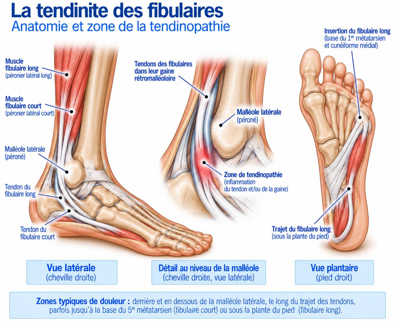
🔍 Click on the image to enlarge it
Function of the fibularis muscles
The fibularis muscles play several roles: they produce eversion (turning the sole of the foot outwards), contribute to the lateral stabilisation of the ankle — particularly during running, changes of direction, landings and on uneven ground — and help control foot position during the stance phase. Fibularis longus also plays a specific role in forefoot stability and control of the foot arch. The fibularis muscles therefore act as a genuine dynamic line of defence against inversion stresses on the ankle.
Biomechanics
During stance, the fibularis muscles must control the movements of the foot; they are particularly loaded during changes of direction, unstable footing, jump landings, running on uneven ground, descents and accelerations. After a lateral ankle sprain, they are also heavily called upon to stabilise the joint — which explains why fibularis disorders are frequently associated with chronic ankle instability.
Although their functions are similar, fibularis brevis and fibularis longus are not necessarily affected in the same way: fibularis brevis is more often painful behind the malleolus and then towards the base of the fifth metatarsal, and may develop longitudinal splits; fibularis longus may be painful behind the malleolus, lower down on the outer edge of the foot, or sometimes under the foot, and may be involved in painful os peroneum syndrome.
Epidemiology
Fibularis tendinopathy mainly affects runners, trail runners and athletes whose activities involve changes of direction, jump landings or footing on unstable ground — notably skiing, football, basketball and other pivoting sports.
It occurs in two main clinical contexts: progressive overload (increased mileage, hilly terrain, a return to trail running or mountain hiking), or following a lateral ankle sprain, when the pain on the outer side persists even though the sprain itself is improving. Subluxation and dislocation of the fibularis tendons, which are rarer but more severe, particularly affect athletes in pivoting sports, skiers, footballers, basketball players and trail runners.
Chronic fibularis disorders are frequently associated with chronic ankle instability or with certain anatomical features (a shallow fibular groove, cavovarus foot), which sustain a genuine vicious circle of sprain, instability and tendon overload.
Causes & risk factors
Fibularis tendinopathy is most often multifactorial, combining training overload, a history of injury and predisposing biomechanical factors. Three typical scenarios are seen in athletes: progressive overload (for example, increased mileage combined with hilly terrain and trail running, with pain gradually developing behind the malleolus); pain on the outer side that persists after an ankle sprain even though the sprain itself is improving; or symptoms appearing after a change of activity (a return to trail running, mountain hiking, pivoting sports, a large increase in elevation gain). Functionally, severity can be classified into four stages (see below) — a practical classification, but not a universally validated anatomical one.
Training factors
Increased mileage, increased elevation gain, uneven ground, returning after a break, more sessions per week, trail running, speed work.
Traumatic factors
Ankle sprain, inversion injury, jump landing, abrupt change of direction.
Biomechanical factors
Cavovarus foot, lateral ankle instability, poor foot control, weakness of the fibularis muscles.
Chronic ankle instability
Chronic instability can sustain the symptoms: patients often describe their ankle as “giving way easily” or report having had several sprains. The fibularis muscles then have to work harder to stabilise the ankle, which sets up a genuine vicious circle: sprain → instability → fibularis overload → tendinopathy → reduced function → new sprain. Management must therefore address the whole problem, not just the tendon.
Why do the tendons move out of place?
The fibularis tendons pass behind the malleolus in a groove held in place by the superior fibular retinaculum. Damage to this restraining system — most often after a sprain or a sudden, forceful contraction of the fibularis muscles — can allow the tendons to move partially (subluxation) or completely (dislocation) out of their groove. Certain anatomical features favour this: a shallow fibular groove, a particular shape of the malleolus, foot morphology, cavovarus foot, chronic lateral instability.
Symptoms
The pain is generally located on the outer edge of the ankle: behind the lateral malleolus, below the malleolus, along the outer edge of the foot, at the base of the fifth metatarsal, and sometimes under the foot in the case of fibularis longus. It may be described as dull, burning, or as a pulling sensation, mechanical pain or a catching sensation.
It frequently appears during running, changes of direction, on uneven ground, on descents, when jumping or during eversion exercises. Initially present only during or after training, it may later appear when walking, on stairs, at rest, or even at night in the more irritable forms.
Key point
A snapping, clicking or “jumping” tendon sensation behind the malleolus is not simply a symptom of tendinopathy: it should prompt investigation for subluxation or dislocation of the fibularis tendons, an important condition not to miss because it calls for a different treatment approach.
Clinical examination
Inspection looks for swelling, deformity, asymmetry, muscle wasting or any displacement of the tendons. Palpation explores the course of both tendons — fibularis brevis and fibularis longus — from where they pass behind the malleolus to their distal course; localised tenderness that exactly reproduces the usual pain is an important diagnostic finding.
Resisted eversion tests (“turn your foot outwards while I resist”) typically reproduce the pain; it is useful to compare strength, pain and endurance with the opposite side. Stretch tests, combining inversion and dorsiflexion, may also reproduce the pain. Stability assessment includes the anterior drawer test, forced varus and functional tests (single-leg stance, balance, heel raise, single-leg hop, lateral hops, running, change of direction).
When subluxation is suspected, the examination must be dynamic: active dorsiflexion, resisted eversion and rapid ankle movements, looking for snapping, clicking or visible displacement of the tendon. No single test can establish the diagnosis with certainty: it relies on the combination of the clinical history, location, palpation, resisted tests, functional examination and imaging when necessary.
Further tests
Plain X-rays
Look for a fracture, a bony abnormality, an os peroneum, foot morphology or post-traumatic changes. Particularly useful when an associated bony abnormality is suspected.
Ultrasound
Assesses tendon structure, thickening, vascularity, tenosynovitis, tendon split, rupture or subluxation. Its great advantage is that it allows dynamic assessment — particularly valuable when looking for subluxation, snapping or dislocation.
MRI
Assesses the tendons, muscles, ligaments, bone and surrounding tissues. Particularly useful for detecting a longitudinal split, rupture, tenosynovitis, oedema or associated bone pathology. Considered a reference examination for the radiological assessment of fibularis disorders, but the diagnosis remains primarily clinical.
Differential diagnoses
Lateral ankle sprain
Very common and sometimes associated with fibularis involvement — the two can coexist.
Chronic ankle instability
May be the cause or the consequence of the fibularis disorder; each often sustains the other.
Stress fracture
To be considered in the case of very localised bone pain, unlike the more diffuse tendon pain along the course of the fibularis tendons.
Fracture of the base of the fifth metatarsal
A particularly important diagnosis to rule out when pain is attributed to fibularis brevis.
Painful os peroneum syndrome
The os peroneum, a sesamoid bone located within the fibularis longus tendon, can become symptomatic after trauma, overload, fracture or displacement — pain on the lateral edge of the foot or under the cuboid.
Subfibular impingement
Can cause chronic lateral pain close to the course of the fibularis tendons.
Calcaneofibular ligament injury
May be associated with a fibularis disorder, particularly after a sprain.
Sinus tarsi syndrome
Pain that is more anterolateral than the retromalleolar pain typical of the fibularis tendons.
Ankle arthropathy
To be considered in the presence of stiffness and swelling rather than simple mechanical tendon pain.
Classification
Stage 1
Mild irritation: pain after activity, no loss of function, sport almost normal.
Stage 2
Moderate tendinopathy: pain during activity, discomfort when running, reduced performance.
Stage 3
Severe tendinopathy: pain when walking, swelling, loss of strength, instability.
Stage 4
Structural injury: significant tendon split, rupture, subluxation or dislocation of the fibularis tendons.
Treatment
Phase 1 – Reduce irritability and adjust the load
Running, elevation gain, uneven ground, jumping and changes of direction are temporarily reduced — without necessarily stopping all activity. Cycling, swimming, appropriate muscle work and pain-free cardio can be maintained.
In very painful forms, relative and temporary immobilisation may be used depending on severity: a more stable shoe, an ankle brace, or even a walking boot. It should remain time-limited and is mainly useful when pain prevents any normal loading.
Phase 2 – Activating the fibularis muscles
Start with simple exercises: eversion without resistance (3 × 15–20), then eversion with a resistance band, progressing from light to heavier resistance. Isometrics — holding the foot in eversion against resistance, for example 4 × 30–45 seconds — complete this work.
The aim is to gradually restore the contractile capacity of the fibularis muscles without irritating the tendon.
Phase 3 – Progressive strengthening
The load is increased gradually: eversion with a resistance band, eversion with a cable pulley, weight-bearing work, single-leg work, and control of eversion during functional movements. Maximal strength, endurance and speed of contraction are developed progressively.
Phase 4 – Global ankle rehabilitation
The fibularis muscles should not be rehabilitated in isolation: the triceps surae, tibialis anterior, tibialis posterior and intrinsic foot muscles are also strengthened, using exercises such as heel raises, single-leg work, balance and ankle control.
Phase 5 – Proprioception
This phase is essential when there is a history of sprain. It uses single-leg balance, an unstable cushion, manual perturbations, jump landings, exercises with eyes open then closed, and dynamic work.
The aim is to restore the ankle's ability to react quickly to unexpected movements.
Phase 6 – Dynamic work and gradual return to sport
Once strength is sufficient, vertical jumps, lateral jumps, bounds, changes of direction, accelerations and decelerations are introduced, to gradually expose the fibularis muscles to rapid contractions.
For a runner, the following are gradually reintroduced: easy running, flat ground, duration, volume, pace variations, elevation gain, uneven ground and then trail running. For an athlete in a pivoting sport: acceleration, braking, change of direction, lateral movement, jumps.
Before returning to running, several criteria are checked: little or no pain, with no worsening the next day; good ankle mobility without restriction; good eversion, good calf strength and good endurance; stable single-leg stance, with no giving way or subluxation; ability to perform heel raises, jumps, lateral jumps and single-leg landings.
Progression can then follow: walking → jogging → running → accelerations → hills → jumps → changes of direction → uneven ground → sport-specific work. For trail running: flat → gentle slope → controlled descent → technical terrain → full elevation gain. For pivoting sports: running → acceleration → braking → lateral movement → change of direction → contact with opponents.
Yes, you can run with fibularis tendinopathy in some mild forms, provided the pain remains low, does not worsen during the session, your running technique remains normal, symptoms return to their usual level after exercise, and there is no significant swelling. However, high-risk activities must be stopped in the event of significant pain, weakness, tendon snapping, a sensation of dislocation or suspected rupture.
Complementary treatments
Suitable footwear
A stable shoe, good heel support, a sole suited to the terrain, possibly stiffer if the tendon is very irritable. A temporary aid, not a universal treatment.
Ankle brace
Useful after a sprain, in cases of instability or during the return to sport: reduces excessive ankle movement. Should remain a temporary aid and replaces neither strengthening nor proprioceptive training.
Orthotic insoles
To be considered in the case of cavovarus foot, marked supination or a biomechanical abnormality clearly correlated with the symptoms. Not systematic.
Manual therapy
Can improve ankle mobility, temporarily reduce pain, treat certain joint restrictions and facilitate exercises. Does not replace strengthening.
Shockwave therapy
Limited data specific to fibularis tendinopathies; not enough evidence to date to make it a first-line treatment. May be considered in resistant chronic forms, with a low level of evidence.
Corticosteroid injection
To be used with caution. Injection directly into the tendon is generally not favoured because of the risk of weakening the tendon. Injection into the sheath may sometimes be considered in selected cases of tenosynovitis, without replacing rehabilitation.
PRP (platelet-rich plasma)
Very limited data; no evidence strong enough to recommend it routinely. May be considered in some refractory chronic forms, as an individualised decision.
Surgery
Surgery may be considered in cases of rupture, a significant tendon split, recurrent dislocation, persistent symptomatic subluxation, chronic pain despite well-conducted conservative treatment, or an anatomical abnormality favouring recurrence. The procedure depends entirely on the lesion: synovectomy, debridement, tendon repair, tubularisation, retinaculum repair, deepening of the retromalleolar groove, or transfer/reconstruction in certain ruptures. International consensus stresses that surgical treatment should be tailored to the precise pathology rather than applied uniformly.
Tendon splits
A limited split can sometimes be treated by debridement, repair or tubularisation. When tendon loss is substantial, reconstruction or transfer may be necessary. The decision depends on the size of the lesion, the tendon involved, the condition of the tissue and the patient’s activity level.
Subluxation and dislocation
When the tendons sublux or dislocate in a symptomatic and recurrent way, treatment may combine repair of the superior retinaculum, deepening of the fibular groove and treatment of associated abnormalities. Available data show good results after surgery, particularly when retinaculum repair is combined with groove deepening in appropriate indications.
In some acute cases of dislocation, conservative treatment (immobilisation, reduction, maintaining the position, progressive rehabilitation) may be tried, but results are variable: a systematic review found high recurrence rates with strapping alone, whereas prolonged non-weight-bearing immobilisation appeared more effective in the small series available. In an athlete with recurrent dislocation, surgery is far more often considered.
Rehabilitation after surgery
Recovery depends on the procedure performed and generally follows five phases: protection (immobilisation, oedema control, protection of the repair, gradual resumption of weight-bearing as instructed); gradual recovery of ankle mobility; strengthening (fibularis muscles, triceps surae, tibialis anterior, intrinsic foot muscles); proprioception (balance, single-leg stance, perturbations, dynamic work); then reintroduction of impact (jumps, bounds, running, changes of direction).
Recovery time
A mild form of fibularis tendinopathy can improve within a few weeks. Chronic tendinopathy, a tendon split or associated instability, on the other hand, may require several months of treatment.
Return to sport should never be based solely on the disappearance of pain, but on meeting specific return criteria: pain, mobility, strength, stability and function.
Prevention
Preventing recurrence relies mainly on strengthening the fibularis muscles, proprioceptive training, single-leg work, ankle stability, complete rehabilitation after a sprain and a sensible progression of training volume.
After a sprain, returning to running simply because “I can walk without pain” is not enough: you also need to regain strength, balance, control and the ability to absorb stress before resuming full training.
Frequently asked questions
Is it the same thing as peroneal tendinopathy?
Where is the pain located?
Can you have tendinopathy without having had a sprain?
Can an old sprain be responsible?
Why does my ankle snap?
Do I need to stop running?
Should the fibularis muscles be strengthened after a sprain?
Are insoles necessary?
Is surgery common?
How long does it take to heal?
Common mistakes
Treating all lateral ankle pain as a simple sprain, without considering associated or isolated fibularis involvement.
Ignoring a snapping or clicking sensation, which may reveal subluxation of the tendons.
Relying on a static MRI when dynamic subluxation is better detected by dynamic ultrasound.
Strengthening only the fibularis muscles without restoring overall ankle function.
Overlooking chronic ankle instability, which can sustain tendon overload.
Returning directly to technical terrain, even though trail running imposes far greater stresses than easy running on flat ground.
Immobilising an acute form for too long, without gradual reconditioning afterwards.
Injecting directly into the tendon, when the benefit is uncertain and the risk to the tendon is real.
Key points — Institut Basque du Coureur
Fibularis tendinopathy is an important cause of pain on the outer edge of the ankle, particularly in runners, trail runners and athletes whose activities involve changes of direction. It can involve fibularis brevis, fibularis longus, their sheath or their insertion, but above all it is important to distinguish simple tendinopathy from tenosynovitis, a tendon split or rupture, or subluxation or dislocation of the tendons. Diagnosis relies mainly on the clinical history, palpation, resisted tests, stability assessment and functional tests. Ultrasound is particularly valuable for its dynamic nature when subluxation is suspected, while MRI is useful for looking for structural tendon lesions. Treatment of simple tendinopathy relies mainly on load management, progressive strengthening, proprioception, restoring ankle stability and a gradual return to sport. For runners, the goal is not simply to eliminate pain: the aim is to rebuild an ankle that is strong, stable and responsive enough to tolerate the stresses of running, elevation gain and uneven ground. Finally, snapping or a sensation of the tendon "jumping" behind the malleolus should prompt investigation for subluxation or dislocation, a condition that calls for a different treatment approach from simple tendinopathy.
- van Dijk PA, Miller D, Calder J, et al. The ESSKA-AFAS international consensus statement on peroneal tendon pathologies. Knee Surg Sports Traumatol Arthrosc. 2018;26(10):3096-3107.
- Melamed E, Sabo MT. Pathologies of the Peroneals: A Review. Foot Ankle Clin. 2020.
- Peroneal tendon pathologies: From the diagnosis to treatment. Oper Orthop Traumatol. 2021.
- Danna C, Brodell JD Jr, Patel S, et al. Chronic Disorders of the Peroneal Tendons: Current Concepts Review of the Literature. J Am Acad Orthop Surg. 2019;27(16):e752-e760.
- van Dijk PA, Gianakos AL, Kerkhoffs GMMJ, Kennedy JG. Return to sports and clinical outcomes in patients treated for peroneal tendon dislocation: a systematic review. Knee Surg Sports Traumatol Arthrosc. 2016;24(4):1155-1164.
- Non-operative treatment of peroneal tendon dislocations: a systematic review. J Orthop. 2019/2020.
- Peroneal Tendinosis and Subluxation. Clin Podiatr Med Surg. 2020.
- Peroneal tendon injuries: current concepts in diagnosis and treatment.
- Peroneal tendoscopy for peroneal tendon disorders: a systematic review of indications, diagnostic utility, and clinical outcomes. J Orthop. 2025.
- Current trends in tendinopathy: consensus of the ESSKA basic science committee. Part I: biology, biomechanics, anatomy and an exercise-based approach. Knee Surg Sports Traumatol Arthrosc.
Physiotherapy protocol for this injury
Découvrez notre protocole de rééducation détaillé pour peroneal (fibularis) tendinopathy : exercices adaptés, progression structurée et modalités de reprise.
Similar injuries
Retrocalcaneal bursitis
Inflammation of the bursa between the Achilles tendon and the calcaneus, causing pain at the back of the heel in runners.
Ankle sprain
Injury to the ankle ligaments caused by twisting the foot; the most common macrotraumatic injury in runners and athletes.
Ankle impingement syndrome
Compression of the ankle joint tissues during extreme movements.