
Definition
An ankle sprain is an injury to one or more of the ligaments that stabilise the ankle joint. In athletes, and in runners in particular, the most common form is the lateral ankle sprain, usually caused by a sudden inversion movement of the foot combined with internal rotation. It mainly involves the anterior talofibular ligament (ATFL), the calcaneofibular ligament (CFL) and, more rarely, the posterior talofibular ligament (PTFL).
But not all ankle sprains are lateral sprains: medial sprains, tibiofibular syndesmosis sprains, osteochondral lesions, associated fractures and tendon injuries must also be recognised. A “sprain” is therefore a diagnosis of ligament injury, but its location and severity must be precisely determined.
Ankle sprain is one of the most common sports injuries and is often considered minor. Yet a significant proportion of athletes are left, after a first sprain, with a feeling of instability, reduced mobility, loss of strength, proprioceptive deficits, apprehension or recurrent episodes: an inadequately rehabilitated sprain can thus progress to chronic ankle instability. The problem is therefore not only to make the pain go away, but to regain an ankle that can bear load, control, react, accelerate, change direction and run on uneven ground.
Anatomy
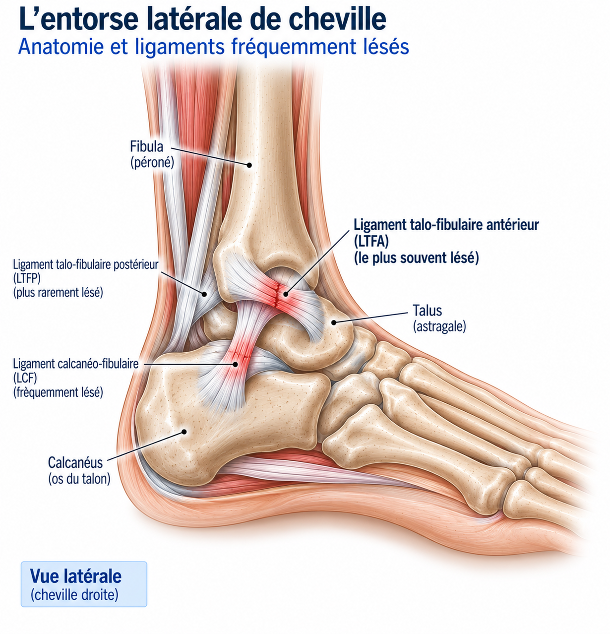
The ankle is made up mainly of the lower end of the tibia, the lower end of the fibula and the talus. The tibia and fibula form a true clamp around the talus, the tibiofibular mortise, which mainly allows dorsiflexion and plantar flexion movements — although ankle function cannot be separated from that of the subtalar joint, the midfoot and the foot.
🔍 Click on the image to enlarge it
The lateral ligaments
The lateral ligament complex comprises three main ligaments. The anterior talofibular ligament is the most frequently injured, being particularly stressed during inversion, plantar flexion and rapid foot movements. The calcaneofibular ligament contributes more to lateral stability when the foot is in a neutral position and is often involved in more severe sprains. The posterior talofibular ligament is much stronger and rarely injured in isolation; its rupture generally indicates a severe sprain.
The medial ligament and the syndesmosis
The medial ligament, or deltoid ligament, stabilises the inner side of the ankle. Much more robust than the lateral ligaments, it is injured less often, but medial sprains should be taken seriously because they may be associated with a fracture, a syndesmosis injury or significant instability. The tibiofibular syndesmosis — the anterior and posterior inferior tibiofibular ligaments and the interosseous membrane — maintains the relationship between the tibia and fibula above the ankle; an injury to it is a syndesmosis sprain (“high ankle sprain”), which generally takes longer to recover from than a simple lateral sprain.
Classic mechanism
The classic mechanism of a lateral sprain combines inversion, internal rotation and plantar flexion: the foot suddenly turns inwards while the body weight continues to move forward, placing particular stress on the anterior talofibular ligament. This mechanism can occur on landing, when running on uneven ground, during a change of direction, or after placing the foot on the outer edge of an irregularity in the ground. In road runners, sprains often occur on an uneven patch, after a misstep or when very fatigued; in trail runners, the situations are more varied (roots, stones, side slopes, technical descents, wet ground, neuromuscular fatigue) — the risk increasing as the ability to react quickly decreases.
After a first sprain, several factors may persist — ligament laxity, proprioceptive deficit, reduced peroneal strength, reduced mobility, impaired postural control, apprehension — making the athlete more vulnerable to a new perturbation: sprain → deficit → instability → new sprain → worsened deficit.
Epidemiology
Ankle sprain is one of the most common sports injuries across all sports, and frequently affects both road runners and trail runners. A significant proportion of athletes are left with residual instability after a first sprain, which explains how common recurrences and chronic ankle instability are in the absence of appropriate rehabilitation.
Causes & risk factors
The classic mechanism of a lateral sprain combines inversion, internal rotation and plantar flexion, usually occurring on landing, when running on uneven ground or during a change of direction.
Risk factors for recurrence
After a first sprain, residual ligament laxity, a proprioceptive deficit, reduced peroneal strength, reduced mobility, impaired postural control or apprehension may persist and predispose to a further sprain. Patients often return to sport as soon as the pain disappears, even though they may still have deficits in strength, balance, mobility or reaction: the disappearance of pain is therefore only one stage of recovery.
Terrain and fatigue factors
In road runners, sprains often occur on an uneven patch of ground, after a misstep or when very fatigued. In trail runners, the situations are more varied: roots, stones, side slopes, technical descents, wet ground. Neuromuscular fatigue reduces control capacity and increases the risk of a wrong movement.
Symptoms
Symptoms depend on severity and generally include pain, swelling, difficulty walking, bruising, limited mobility and a feeling of instability. Pain is usually greatest around the injured ligament; in a lateral sprain, it is located around and slightly in front of the lateral malleolus.
Swelling often appears quickly and may be localised or diffuse; very rapid and massive swelling may indicate intra-articular bleeding or a significant injury, but its extent alone does not allow the severity of the ligament injury to be precisely determined. Bruising may appear in the following hours or days and gradually spread down towards the outer edge of the foot, the heel or the sole, without this migration meaning that the injury is getting worse. In a significant sprain, weight-bearing can be very painful, with limping or walking on the inner edge of the foot; an inability to walk should prompt a search for a fracture, in particular using the Ottawa rules (malleolar pain combined with tenderness on palpation of the posterior edge or tip of the malleolus, or inability to take four steps both immediately after the injury and at the time of assessment).
Clinical examination
The examination should answer several questions: is there a fracture? which ligament is probably injured? is there instability or an associated injury? what is the patient’s functional level and when will they be able to return to running? Systematic palpation looks for tenderness over the anterior talofibular ligament, the calcaneofibular ligament, the malleoli, the base of the 5th metatarsal, the navicular, the syndesmosis and the talus — bone tenderness should raise the possibility of a fracture.
The anterior drawer test mainly assesses the stability of the anterior talofibular ligament (forward translation of the talus relative to the tibia) and is generally more informative a few days after the injury, once the acute pain and swelling subside. The talar tilt test (forced varus) assesses lateral stability, in particular the calcaneofibular ligament. Pain above the joint should prompt a search for syndesmosis involvement (squeeze test, external rotation stress test). Examination of the peroneal tendons (pain behind the lateral malleolus, pain on resisted eversion, weakness, subluxation) and assessment of dorsiflexion (Weight-Bearing Lunge Test) complete the examination. Once the acute phase has passed, functional assessment (single-leg balance, calf raise, squat, jumping, running, changes of direction) allows a gradual shift from anatomical examination to function and then to sport.
Further tests
X-ray
Indicated according to the Ottawa rules when a fracture is suspected (malleolar bone tenderness, inability to take four steps) — these rules help to limit unnecessary X-rays while maintaining excellent sensitivity for fractures.
Ultrasound
Useful for examining ligaments, tendons and superficial lesions, with the possibility of dynamic assessment.
MRI
Particularly useful in cases of persistent pain, suspected osteochondral lesion of the talus, syndesmotic injury, tendon injury, atypical symptoms or failure of management: it can reveal a ligament rupture, bone oedema, an osteochondral lesion, involvement of the peroneal tendons or the deltoid ligament, synovitis or an effusion. Mainly useful when the clinical course is not as expected.
Differential diagnoses
Fracture (malleolar, talus, base of the 5th metatarsal)
Must be systematically ruled out using the Ottawa rules: bone tenderness on malleolar palpation, or inability to take four steps immediately after the injury.
Syndesmosis sprain ("high ankle sprain")
Pain above the joint, pain on external rotation and on tibiofibular compression; recovery generally takes longer than for a simple lateral sprain.
Medial sprain (deltoid ligament)
Much less common than a lateral sprain; significant pain on the inner side should prompt a search for an associated fracture, syndesmosis injury or osteochondral lesion.
Osteochondral lesion of the talus
To be suspected in the presence of persistent pain, recurrent swelling, locking or catching sensations after the sprain; MRI may be useful.
Peroneal tendon injury
Pain behind the lateral malleolus, pain on resisted eversion, weakness or subluxation; may be associated with the sprain or explain persistent pain after it.
Subtalar instability
To be considered in the presence of persistent instability that is poorly explained by the talofibular ligaments alone.
Classification
Grade I
Ligament stretching, moderate pain, limited swelling, stability largely preserved, weight-bearing possible.
Grade II
Partial ligament rupture, more significant pain, significant swelling, limited weight-bearing, moderate laxity.
Grade III
Complete ligament rupture, major swelling, frequent bruising, instability, major difficulty with or inability to bear weight.
Treatment
Acute phase (48-72 h) — control and early weight-bearing
Modern treatment is based on relative protection, gradual weight-bearing, functional rehabilitation and prevention of recurrence — the aim is generally not to immobilise the ankle completely for several weeks. In the first 48 to 72 hours, the aim is to control pain, swelling and difficulty walking: compression, elevation, ice (a symptomatic treatment, with no proven effect on ligament healing itself), a semi-rigid brace and crutches if necessary — rest should remain relative. Early weight-bearing is generally preferable to prolonged immobilisation when the severity of the injury allows: it stimulates proprioception, strength, mobility and confidence in the ankle, with the load kept adapted to pain and stability. Crutches should be gradually reduced as soon as walking becomes possible.
Restoring mobility and isometric work
As soon as possible, dorsiflexion, plantar flexion, controlled ankle movements and toe mobility are worked on (active movements, tracing the alphabet with the foot, gentle dorsiflexion mobilisation). Isometric contractions (eversion, inversion, plantar flexion, dorsiflexion against resistance) stimulate the muscles without significant movement, which is particularly useful when dynamic movements are still painful.
Strengthening the peroneals, calf and foot
The peroneals, which are essential for lateral stability, are strengthened gradually (eversion with a resistance band, weight-bearing eversion, single-leg work, fast and then reactive), progressing from slow to fast and from simple to functional. The triceps surae is strengthened at the same time (double-leg then single-leg calf raises, with the knee straight and bent, weighted work), as it contributes strongly to stability and propulsion during running. The intrinsic foot muscles, which play an active role in balance control, are also worked on (short foot, toe flexion, arch holding, single-leg exercises).
Proprioception
Proprioception is a central part of rehabilitation, because after a sprain the ability to quickly detect and correct foot inversion may be reduced. The progression is: single-leg balance → balance with upper limb movements → unstable surface → eyes closed → external perturbations → fast, unpredictable reactions, in order to restore the perception of foot position, activation of the stabilising muscles, reaction speed and postural control.
Jumps, changes of direction and reactive work
Once strength and balance have been sufficiently regained, double-leg then single-leg jumps, forward-backward and lateral jumps, and landings are introduced — the quality of the landing being as important as the height of the jump. The runner then gradually learns to control braking, acceleration, turning and lateral movement, progressively increasing speed. Finally, the work becomes unpredictable (changing direction on a signal, landing after moving, reacting to a visual or auditory stimulus) in order to reproduce the demands of a real sporting situation.
Gradual return to running
Return to running can begin when walking is comfortable, mobility is sufficient, calf exercises are well tolerated, single-leg balance is satisfactory and small jumps are possible. Start on flat ground, at a slow pace and for a short duration, for example alternating 2 minutes of running with 1 minute of walking, then gradually increase running time, volume and speed, before reintroducing changes of direction and then the uneven terrain specific to the sport. For trail runners, the progression goes road → compacted path → uneven path → side slopes → descents → technical terrain, with technical descents often among the last stages to be reintroduced.
Complementary treatments
Ankle brace
Particularly useful when returning to a sport with a risk of recurrence: it limits certain dangerous movements, improves confidence and reduces the risk of a new sprain. However, it should not replace rehabilitation.
Taping
Additional protection, particularly useful for athletes with residual instability or returning quickly. Temporary effect.
Ice
May temporarily reduce pain, the sensation of heat and discomfort, but should not be regarded as a treatment that on its own speeds up ligament healing: its value is mainly symptomatic.
Surgery
The vast majority of acute lateral sprains are treated without surgery; a complete ligament rupture visible on MRI (for example of the anterior talofibular ligament) is not in itself a systematic indication — surgery should not be decided on this basis alone. It may be discussed in cases of persistent chronic instability, failure of properly conducted rehabilitation, a significant ligament injury with instability, an osteochondral lesion requiring treatment, certain syndesmosis injuries or particular associated injuries.
In some cases of chronic instability, reconstruction or repair of the lateral ligaments may be offered, aiming to restore mechanical stability, confidence and function — but it should generally be combined with a full postoperative rehabilitation programme.
Recovery time
Recovery time depends enormously on severity: a mild sprain may require a few weeks to return to normal activity, a moderate sprain often requires several weeks of sports recovery, a severe sprain may require several weeks to several months, and a syndesmosis sprain can take a particularly long time to recover. These timeframes are only a guide: functional recovery matters more than the number of days elapsed, and two runners with a sprain of similar grade can progress very differently.
Return to sport follows the international PAASS framework, which recommends assessing five domains rather than time elapsed alone: Pain (pain during sport and over the last 24 hours), Ankle impairments (mobility, strength, endurance, power), Athlete perception (confidence, feeling of stability), Sensorimotor control (proprioception, dynamic balance) and Sport/functional performance (jumps, agility, sport-specific movements, full training). Persistent pain after several weeks is not necessarily abnormal — ligaments heal gradually, and swelling, mobility, strength or proprioception may remain impaired for some time — but a lack of gradual improvement should lead to the diagnosis being reassessed.
Prevention
Preventing recurrence should be considered an integral part of treatment. It is based on strengthening (peroneals, triceps surae, intrinsic foot muscles, proximal chain), proprioception (single-leg balance, perturbations, jumps, fast reactions) and sport-specific work (changes of direction, uneven terrain, side slopes, descents) — proprioceptive and neuromuscular exercise programmes playing an important role in preventing recurrence. Managing fatigue is also essential, as it reduces neuromuscular control and increases the risk of a wrong movement.
For trail runners, it is particularly useful to work on single-leg balance, lateral jumps, landing, moving over uneven terrain and control on descents, so that the foot is able to react even before the runner is aware of the unevenness of the ground.
Frequently asked questions
Can I walk with a sprain?
Can I keep running?
How long should I wear an ankle brace?
Do I need an MRI?
Can a sprain heal in a week?
Can you have a sprain without swelling?
Do ligaments grow back?
Does a complete ligament rupture need surgery?
Why does my ankle still give way several months later?
Does an ankle brace stop the muscles from working?
Common mistakes
"It's only a sprain": a sprain can progress to chronic instability if it is inadequately rehabilitated.
Immobilising for too long, which can promote stiffness, weakness and loss of proprioception.
Returning as soon as the pain disappears, even though pain does not necessarily reflect functional recovery.
Doing only balance exercises, without restoring strength, endurance, power and fast reactions.
Going straight back to trail running, whereas technical terrain should be reintroduced gradually.
Forgetting the syndesmosis: high or unusual pain should prompt a search for another ligament location.
Requesting an MRI systematically, even though it is not necessary for all sprains.
Relying solely on the anatomical grade, which is not enough to determine return to sport.
Key points — Institut Basque du Coureur
Ankle sprain is one of the most common injuries in athletes. In runners, it is most often lateral, following a sudden inversion movement, but ankle pain after an injury should never automatically be regarded as a simple lateral sprain: a fracture, a syndesmosis sprain, a medial sprain, a peroneal injury, an osteochondral lesion or subtalar instability must be looked for. The Ottawa rules help to select the patients who need an X-ray. Prolonged immobilisation is generally not necessary in simple lateral sprains: gradual weight-bearing and mobilisation are important, and rehabilitation should combine mobility, strength and proprioception, with strengthening of the peroneals. Jumps and changes of direction should be reintroduced before a full return to sport, and uneven terrain should be reintroduced gradually in runners, and particularly in trail runners. An ankle brace can be useful when returning to sport. The disappearance of pain is not enough to allow a return to sport: this must take into account pain, ankle impairments, the athlete's confidence, sensorimotor control and functional performance. A well-treated ankle sprain is not simply an ankle that no longer hurts: it is an ankle that can react quickly, withstand stress and remain stable when the ground does not behave as expected.
- Gribble PA, Bleakley CM, Caulfield BM, et al. 2016 consensus statement of the International Ankle Consortium: prevalence, impact and long-term consequences of lateral ankle sprains. Br J Sports Med. 2016;50:1493-1495.
- Doherty C, Bleakley C, Hertel J, et al. Recovery from a first-time lateral ankle sprain and the predictors of chronic ankle instability: a prospective cohort analysis. Am J Sports Med. 2016;44:995-1003.
- Tassignon B, Verschueren J, Delahunt E, et al. Criteria-based return to sport decision-making following lateral ankle sprain injury: a systematic review and narrative synthesis. Sports Med. 2019;49:601-619.
- Smith MD, Vicenzino B, Bahr R, et al. Return to sport decisions after an acute lateral ankle sprain injury: introducing the PAASS framework — an international multidisciplinary consensus. Br J Sports Med. 2021;55:1270-1276.
- Hertel J, Corbett RO. An updated model of chronic ankle instability. J Athl Train. 2019;54:572-588.
- Martin RL, Davenport TE, Paulseth S, et al. Ankle stability and movement coordination impairments: lateral ankle ligament sprains revision 2021. J Orthop Sports Phys Ther. 2021.
- Vuurberg G, Hoorntje A, Wink LM, et al. Diagnosis, treatment and prevention of ankle sprains: update of an evidence-based clinical guideline. Br J Sports Med. 2018;52:956.
- Picot B, et al. Ankle-GO score is associated with the probability of becoming coper after lateral ankle sprain: a 1-year prospective cohort study. Br J Sports Med. 2024;58:1115-1122.
- American College of Radiology. ACR Appropriateness Criteria®: Acute Trauma to the Ankle. 2026.
Similar injuries
Achilles tendinopathy
Inflammation of the Achilles tendon causing pain and stiffness at the heel, the most common injury in runners.
Blisters in runners
Fluid-filled pockets under the skin caused by friction, heat and moisture, common in running, trail running and marathons.
Tibialis posterior tendinopathy
Pain on the inner side of the ankle caused by irritation of the tendon that stabilises the arch of the foot.