
Definition
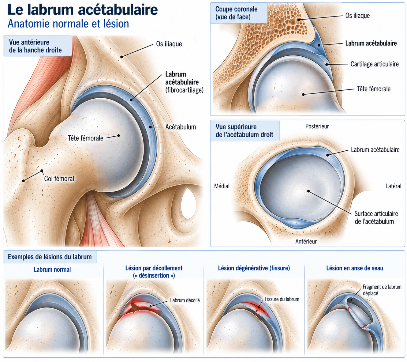
The acetabular labrum is a fibrocartilaginous ring surrounding the rim of the acetabulum, the socket of the pelvis in which the femoral head articulates. A hip labral tear refers to a tear, detachment, rupture or degeneration of this tissue.
In runners, it can cause groin or hip pain, but its mere presence on imaging does not necessarily mean that it is responsible for the symptoms: labral tears are frequently seen in people with few or no symptoms.
Labral tears are frequently associated with femoroacetabular impingement (FAI/FAIS), a cam or pincer hip morphology, hip dysplasia, repetitive sports overload, trauma or a twisting movement, or associated cartilage lesions. Moreover, the diagnosis of femoroacetabular impingement syndrome is not based solely on the presence of a bony morphology or a labral tear: it requires agreement between symptoms, clinical signs and imaging.
Anatomy
The labrum is a fibrocartilaginous ring attached to the rim of the acetabulum, the socket of the pelvis that receives the femoral head. It plays several essential roles in hip mechanics.
Deepening the acetabulum
It increases the functional depth of the acetabular socket and thus contributes to hip stability.
“Seal” effect
The labrum helps maintain a suction effect around the femoral head, a function that also contributes to joint stability.
Load distribution
It helps distribute pressure between the femoral head and the acetabulum.
Cartilage protection
It helps protect the articular cartilage and maintain the mechanical properties of the synovial fluid. A labral tear can therefore alter joint mechanics, but its clinical significance depends heavily on the context: bony morphology, state of the cartilage, and associated symptoms and clinical signs.
🔍 Click on the image to enlarge it
The different types of labral tears
A tear may be peripheral, longitudinal, radial, complex, or detached from the acetabular rim, with or without associated cartilage damage. It may also occur in isolation, or be associated with cam impingement, pincer impingement, dysplasia or early osteoarthritis. This distinction matters: a labral tear associated with dysplasia is not treated in the same way as a tear related to cam impingement.
Epidemiology
Labral tears mainly affect runners, pivoting sports athletes (football, tennis, dance), people practising sports involving deep flexion, and young active adults (20–45 years). Many tears are present without pain but become symptomatic during mechanical overload. In runners, they are a common cause of persistent groin pain, unexplained discomfort on returning to sport, and chronic hip pain.
Causes & risk factors
Mechanisms of labral injury
The labrum can be subjected to significant stress during movements combining hip flexion, adduction, internal rotation, external rotation and axial loading. Repeated movements at end range can cause repetitive stress on the acetabular rim. In athletes, labral tears are particularly common when the hip has a morphology that predisposes to femoroacetabular impingement.
Femoroacetabular impingement (FAI)
Femoroacetabular impingement, or FAI, is a common cause of labral tears in athletes. There are two main morphologies, which can also occur together (mixed form).
Cam morphology: there is a bony prominence at the femoral head–neck junction. During hip flexion and rotation, this prominence can impinge on the acetabular rim.
Pincer morphology: the acetabular rim covers more of the femoral head. Certain positions can then compress the labrum against the femoral neck.
Key point
A cam or pincer morphology is not necessarily pathological: many athletes have a cam-type morphology without any pain. Femoroacetabular impingement syndrome means the presence of a compatible morphology combined with consistent symptoms and clinical signs — not merely the presence of an imaging finding.
Why are runners affected?
Running involves thousands of cycles of flexion-extension, loading, pelvic rotation and single-leg stabilisation: the hip must control the movement of the pelvis with every foot strike. Certain situations can increase the stresses: a sudden increase in training volume, trail running, significant elevation changes, speed sessions, a long stride, hill work, muscle fatigue or a major change in running technique.
However, there is no evidence that running alone is responsible for a labral tear: hip morphology, medical history, sporting demands and individual factors all interact.
Symptoms
The most typical presentation is groin pain: the patient often describes a deep, anterior pain located in the groin. They may show the painful area by cupping the front of the hip with their hand — a sign sometimes called the C-sign.
Positions combining marked flexion and rotation can provoke symptoms: deep squatting, crouching, getting in and out of a car, climbing certain stairs, prolonged sitting. During a run, pain may appear at the start of the session, after several kilometres, during accelerations, on hills or after the session. Some patients also experience clicking, snapping, a catching sensation or a feeling of locking — although these symptoms are not specific to the labrum.
In runners, the pain can be difficult to locate: it may be felt in the groin, at the front of the hip, in the buttock area, on the side of the hip, or sometimes in the thigh. Clearly lateral pain should prompt a search for gluteal tendinopathy or trochanteric bursopathy; posterior pain should prompt a search for other diagnoses.
A labral tear can also cause discomfort in flexion, pain during rotation, reduced performance or limitation of certain ranges of motion. However, a genuinely marked limitation of mobility should prompt a search for more widespread joint involvement.
Clinical examination
The examination starts with an overall assessment of the lower limb: walking, running if possible, hip mobility, muscle strength, pelvic control, single-leg stability, and mobility of the lumbar spine, knee, sacroiliac joint and peritrochanteric structures.
Hip mobility is assessed in all planes (flexion, extension, abduction, adduction, internal rotation, external rotation). Limited internal rotation, particularly in flexion, may be found in femoroacetabular impingement syndrome — but no single limitation is enough on its own to make the diagnosis.
Several provocation tests can be used. The FADIR test (flexion, adduction, internal rotation) is frequently positive in femoroacetabular impingement syndrome; its high sensitivity mainly means that a negative test can be useful to rule out certain conditions, but a positive test is not specific to a labral tear. The FABER test (flexion, abduction, external rotation) explores different structures of the hip and looks for provoked pain. The log roll test (passive rotation of the lower limb) can reproduce intra-articular pain or reveal laxity. The resisted flexion test can reproduce some anterior hip pain. However, no single test can diagnose a labral tear on its own.
Further tests
X-rays
Essential when a labral tear or femoroacetabular impingement is suspected. The 2025 international consensus recommends at least a weight-bearing anteroposterior pelvis view and a 45° Dunn-type view, to look for cam morphology, pincer morphology, dysplasia, joint space narrowing, osteophytes or other bony abnormalities.
MRI
Allows assessment of the labrum, cartilage, subchondral bone, muscles, tendons and periarticular structures. It can confirm a labral tear and look for associated lesions. Note, however: a labral abnormality on MRI is not synonymous with disease, as labral tears can be seen in people with few or no symptoms — hence the need to look for agreement between symptoms, clinical examination and imaging before any decision, particularly a surgical one.
MR arthrography
Intra-articular injection of contrast medium before MRI can improve visualisation of certain labral tears, but is not systematically necessary with modern MRI techniques. The choice between MRI and MR arthrography depends on the quality of the scan available, the clinical question and the radiologist's experience.
Diagnostic injection
When there is doubt about the origin of the pain, an intra-articular hip injection (local anaesthetic) can have diagnostic value: temporary disappearance of pain when walking, running or performing a usually painful movement points to an intra-articular origin. The 2025 consensus recommends considering this injection when the intra-articular origin of the pain remains uncertain; however, it should not be regarded as absolute proof and must be interpreted together with all the clinical data.
Differential diagnoses
Femoroacetabular impingement
Very frequently associated with labral tears; requires agreement between symptoms, clinical examination and imaging rather than the mere presence of a bony morphology.
Adductor tendinopathy
Groin pain, particularly during resisted adduction movements.
Psoas tendinopathy
Anterior hip pain, sometimes with snapping (psoas snapping).
Gluteal tendinopathy
Lateral hip pain, rather than groin pain.
Greater trochanteric pain syndrome
Lateral pain, typically reproduced in single-leg stance.
Muscle injury
Particularly of the adductors, rectus femoris or iliopsoas.
Pubalgia
Groin pain that may be multifactorial, often related to efforts such as changes of direction or kicking.
Osteitis pubis
Pubic and groin pain, to be distinguished from an intra-articular origin.
Lumbar spine disorders
Radicular or referred pain from the lumbar spine can be felt in the hip area.
Stress fracture (femoral neck)
To be specifically ruled out in runners with progressive, deep pain, sometimes present at rest.
Hip osteoarthritis
To be considered particularly in older patients, especially when there is associated joint space narrowing.
Classification
Grade 1 – Mildly symptomatic tear
Occasional pain, running possible, no significant limitation, no true locking.
Grade 2 – Symptomatic tear
Regular pain when running, discomfort in flexion, reduced training volume, pain after sessions.
Grade 3 – Highly symptomatic tear
Frequent pain, significant limitation of running, pain in daily life, frequent catching, reduced mobility.
Grade 4 – Tear associated with significant structural disease
Significant femoroacetabular impingement, dysplasia, cartilage lesion, osteoarthritis, instability or true locking. Management should then be specialised.
Treatment
Is surgery always necessary?
No: initial treatment can be non-surgical, particularly when the pain remains moderate, there is no true locking or advanced osteoarthritis, and function can be improved through rehabilitation. The ISHA consensus notably recommends management combining education, activity modification and targeted rehabilitation before considering surgery in appropriate situations.
The goal of rehabilitation is to restore strength, pelvic control, dynamic stability, load tolerance and the ability to manage the hip's end ranges of motion.
Managing load and irritating positions
The first step is to identify the activities that reproduce the symptoms. Fast running, hills, downhills, very long strides, deep squats, pivoting movements and exercises requiring deep hip flexion can be temporarily reduced. Running can often be continued if it remains well tolerated: the aim is not to put the hip on complete rest.
In femoroacetabular impingement, certain positions combining flexion, adduction and internal rotation can be particularly symptomatic. It can therefore be helpful to temporarily limit deep flexion, very low seating, deep squats, prolonged cross-legged positions and forced end-range movements — without trying to avoid all these positions permanently. The goal is to gradually rebuild the hip's tolerance to these ranges of motion.
Glute strengthening, then weight-bearing strengthening
Hip control is fundamental for runners. Work begins with glute strengthening: hip abduction (side-lying or with a resistance band), clamshell (external rotators), glute bridge progressing to hip thrust, and lateral band walks for the gluteus medius.
Progression continues towards functional weight-bearing exercises: partial squat, split squat, lunge, step-up, step-down, deadlift and single-leg work, with depth gradually increased according to tolerance. The aim is not to avoid all hip flexion, but to restore sufficient functional capacity.
Pelvic control and trunk work
A runner must be able to control their pelvis on a single leg: single-leg balance, assisted single-leg squat, step-down, single-leg stance with arm movements and dynamic pelvic work help train pelvic stability, knee control, control of hip adduction and trunk–pelvis–lower limb coordination.
Trunk control directly influences hip mechanics: side plank, front plank, Pallof press, anti-rotation work and single-leg exercises improve the transmission of forces between the trunk, pelvis, hip, knee and foot.
Reintroducing impacts and returning to running
Before a full return to running, you can progress in stages: brisk walking, stair climbing, small two-footed hops, single-leg hops, bounding, stride work and then running. The hip's response over the following 24 hours should guide progression.
The return to running can then follow a multi-stage progression: alternating one minute of running with two minutes of walking for 20 to 30 minutes, then increasing running time, short continuous running, gradually increasing distance, reintroducing hills, reintroducing speed and finally returning to usual volume. It is best to change only one parameter at a time.
Criteria for returning to running
Before a regular return, the ideal is to have little or no pain in daily life, functional mobility, sufficient strength, good single-leg stability, no pain during squats, good tolerance of jumping and no significant symptomatic reaction after training.
Progression should then be guided by pain, function, strength, endurance, neuromuscular control and load tolerance — rather than by a fixed timeline.
Complementary treatments
Intra-articular injection (diagnostic ± symptomatic)
May reduce symptoms, but does not repair the labrum. Its value is mainly diagnostic when the intra-articular origin of the pain remains uncertain, by assessing whether the pain temporarily disappears when walking, running or performing a usually painful movement.
Surgery
When should surgery be considered?
Surgery may be discussed when symptoms are persistent, functional impairment is significant, properly conducted rehabilitation has not brought sufficient improvement, the intra-articular diagnosis is well established, a correctable structural lesion has been identified, and the patient has a morphology suitable for joint-preserving surgery.
The decision must be individualised: age, the state of the cartilage, dysplasia, osteoarthritis and the severity of symptoms influence the prognosis. The 2025 consensus notably considers age, pain severity, dysplasia and degenerative changes to be important prognostic factors for the surgical outcome.
Hip arthroscopy
The procedure may involve several steps. When the labrum is repairable, the current aim is, as far as possible, to preserve and repair it; when the tissue is irreparable or insufficient, reconstruction may be considered in certain situations. Osteoplasty can correct the bony prominence responsible for cam impingement, and targeted bone resection can correct an acetabular rim responsible for pincer impingement. The joint capsule must also be taken into account in order to preserve or restore hip stability.
Modern surgery therefore aims to treat the mechanical cause associated with the labral tear, and not simply to remove the labrum.
Rehabilitation after surgery
Recovery depends on the type of tear, the type of repair, any cam/pincer correction or reconstruction, the state of the cartilage and the sporting level. There is no single protocol applicable to all hip arthroscopies: weight-bearing and permitted ranges of motion depend on the surgical procedure performed.
Post-operative rehabilitation is generally organised in several phases: an early phase aimed at controlling pain and inflammation, protecting the repaired tissues and gradually recovering the permitted mobility; a muscle recovery phase (gluteus medius activation, strengthening of the gluteus maximus, rotators, quadriceps and hamstrings, core work, single-leg work) aimed at functional symmetry; an advanced strengthening phase gradually introducing loaded squats, split squats, lunges, deadlifts, step-ups, step-downs, single-leg work and multidirectional exercises; then a power phase (jumps, bounding, plyometrics, accelerations, changes of pace), conditional on prior recovery of strength and motor control. The running-specific phase then follows a progression comparable to that of conservative treatment (brisk walking, walk-run intervals, continuous running, increasing distance, accelerations, hills, specific work, return to usual volume), with speed and hills only reintroduced once good tolerance of easy running has been regained.
Return to sport after surgery
The return should not be determined by the calendar alone. Criteria include mobility, pain, strength, stability, neuromuscular control, endurance, power, agility and sport-specific tasks: modern rehabilitation programmes after hip arthroscopy increasingly use a criteria-based rather than strictly time-based progression.
Published data generally report high rates of return to sport after surgery for femoroacetabular impingement, but returning to the same level depends in particular on the state of the cartilage, the type of sport and the initial level: one systematic review reported approximately 87% return to sport and 82% to the same level, with considerable heterogeneity between studies.
Prognosis
The prognosis depends heavily on the overall condition of the hip. Favourable factors include younger age, moderate pain, preserved cartilage, the absence of significant osteoarthritis or dysplasia, an accurate diagnosis, well-conducted rehabilitation and correction of a mechanical abnormality when indicated. Unfavourable factors include osteoarthritis, significant cartilage damage, dysplasia, severe and long-standing pain, older age and significant degenerative changes. An isolated labral tear in a young runner with preserved cartilage therefore does not have the same prognosis as a labral tear associated with hip osteoarthritis.
Recovery time
Timeframes vary widely. A gradual return to running generally takes place several months after surgery, and for athletes, a full return to sport is often around 6 to 8 months, with considerable variation depending on the type of procedure, the labral repair, the bony correction performed, the sporting level, the recovery of strength and the functional criteria achieved.
It is therefore best to avoid giving the patient a fixed return date: the timeline guides rehabilitation, the criteria determine progression.
Prevention
There is no programme that can prevent all labral tears, but certain strategies can be helpful.
Load progression should avoid sudden increases in mileage, intensity, elevation gain or speed work. Muscle strengthening (glutes, quadriceps, hamstrings, trunk muscles) should be maintained regularly, as should single-leg control: the runner must be able to stabilise their pelvis properly during stance.
Range-of-motion management means avoiding unnecessary repetition of forced end-range movements when they are painful. Finally, varying the stresses by alternating running, cycling, strengthening, mobility and recovery helps limit repetitive overload of the hip.
Frequently asked questions
Can a labral tear heal?
Can you run with a labral tear?
Is a labral tear always painful?
Is femoroacetabular impingement the same as a labral tear?
Is rest enough?
Is cycling allowed?
Can you do squats?
Is an MR arthrogram necessary?
Can a hip injection treat the tear?
When is surgery needed?
Can you get back to your sporting level after surgery?
Common mistakes
Assuming that an MRI showing a tear necessarily explains the pain, without looking for clinical agreement.
Believing that the labrum must absolutely be repaired, when some tears are asymptomatic or respond well to conservative management.
Systematically trying to stretch the hip to the maximum, which can be irritating in femoroacetabular impingement; rehabilitation should prioritise control and muscle capacity rather than the pursuit of maximum range of motion.
Thinking that you have to stop running completely, when a temporary adjustment of load may be enough in some situations.
Believing that after an operation, running will be possible within 6 weeks: a labral repair and/or bony correction generally requires several months of rehabilitation.
Thinking that the labrum will grow back after resection — this is one of the reasons why modern strategies favour preserving and repairing the labrum wherever possible.
Assuming that femoroacetabular impingement is necessarily the cause of the tear, when the relationship between cam/pincer morphology, sporting activity, symptoms and labral tear is complex.
Key points — Institut Basque du Coureur
A hip labral tear does not automatically mean surgery. In runners, it must be interpreted in context, looking in particular for femoroacetabular impingement, cam or pincer morphology, dysplasia, cartilage damage or another cause of groin pain. Diagnosis is based on agreement between symptoms, clinical examination and imaging: a tear visible on MRI can be completely asymptomatic. Initial treatment can often combine load adjustment, temporary modification of irritating positions, strengthening of the hip muscles, trunk work, pelvic control, single-leg rehabilitation and a gradual return to impact activities and running. When surgery is necessary, the modern aim is to preserve and repair the labrum where possible, while correcting the associated mechanical cause when there is one. Finally, in runners, the return to sport should not be decided on a date alone: the hip must first regain sufficient mobility, strength, control, power and load tolerance to withstand the specific demands of running.
- Matache BA, Belzile ÉL, Ayeni OR, et al. Management of Labral Tears in the Hip: A Consensus Statement. Orthop J Sports Med. 2025;13(1):23259671241305409.
- Griffin DR, Dickenson EJ, O'Donnell J, et al. The Warwick Agreement on femoroacetabular impingement syndrome (FAI syndrome): an international consensus statement. Br J Sports Med. 2016;50:1169-1176.
- Casartelli NC, Leunig M, Maffiuletti NA, Nötzli HP. Quantitative assessment of hip muscle strength in femoroacetabular impingement. J Orthop Sports Phys Ther. 2011.
- Wall PDH, Fernandez M, Griffin DR, Foster NE. Nonoperative treatment for femoroacetabular impingement: a systematic review of the literature. PM R. 2013;5:418-426.
- Reiman MP, Peters S, Sylvain J, Hagins M, Liggett C, Goode AP. Femoroacetabular impingement surgery and rehabilitation. J Orthop Sports Phys Ther. 2014.
- Enseki M, Harris-Hayes M, White DM, et al. Nonoperative management of individuals with femoroacetabular impingement: a clinical commentary. J Orthop Sports Phys Ther. 2018.
- Edelstein J, Ranawat A, Enseki KR, Yun RJ, Draovitch P. Post-operative guidelines following hip arthroscopy. Curr Rev Musculoskelet Med. 2012;5:15-23.
- Domb BG, Stake CE, Botser I, Jackson TJ. Surgical dislocation of the hip versus hip arthroscopy for the treatment of femoroacetabular impingement: a systematic review. Arthroscopy. 2013.
- Griffin DR, et al. The UK FASHIoN randomized controlled trial of arthroscopic surgery for hip impingement versus best conservative care. BMJ. 2018.
- Minkara AA, Westermann RW, Rosneck J, Lynch TS. Systematic review and meta-analysis of return to sport after hip arthroscopy for femoroacetabular impingement. Am J Sports Med. 2019.
- Holling M, Miller ST, Geeslin A. Rehabilitation and Return to Sport After Arthroscopic Treatment of Femoroacetabular Impingement. Arthrosc Sports Med Rehabil. 2022;4:e125-e132.
- Gard S, Christofilopoulos P, Ménétrey J. Femoroacetabular impingement: rehabilitation and return to sport. Rev Med Suisse. 2015;11:1450-1455.
- Matta J, et al. Contemporary concepts in hip preservation surgery and return to sport after hip arthroscopy.
Similar injuries
Gluteus medius tendinopathy
Pain on the outside of the hip caused by irritation of the gluteus medius tendon.
Anterolateral TFL impingement
Anterior hip irritation associated with overload of the tensor fasciae latae.
Femoroacetabular impingement
Abnormal contact between the femoral head and the pelvis, causing groin pain.