
Definition
Anterolateral impingement of the tensor fasciae latae (TFL) refers to pain localised to the anterolateral part of the hip or the upper thigh, over the origin of the TFL and the proximal part of the iliotibial band.
It is a condition that is relatively little described in the literature compared with other causes of lateral hip pain. It can, however, be seen in runners, particularly when the TFL is heavily and repeatedly loaded in positions of hip flexion, abduction and internal rotation, or when its role in stabilising the pelvis becomes excessive.
The clinical picture may correspond to several overlapping mechanisms: musculotendinous overload of the TFL, enthesopathy of its iliac origin, irritation of the proximal part of the iliotibial band, mechanical impingement between the TFL, the fascia lata and the anterolateral bony structures, myofascial pain of the TFL or, more rarely, snapping or dynamic displacement of the superficial tissues.
It is therefore better to regard “anterolateral TFL impingement” as a functional clinical diagnosis, rather than as a single, clearly defined anatomical lesion.
Anatomy
The tensor fasciae latae is a small muscle located on the anterolateral part of the hip. It arises from the anterior superior iliac spine (ASIS) and the anterior part of the iliac crest, then quickly continues as the fascia lata, whose fibres largely make up the iliotibial band.
The TFL works in synergy with the gluteus medius, gluteus minimus, gluteus maximus and the iliotibial band. Its main functions are hip flexion, abduction, internal rotation, lateral stabilisation of the pelvis, and a contribution to knee stabilisation via the iliotibial band.
In runners, its role becomes particularly important during phases of dynamic stabilisation and acceleration: the TFL is especially loaded when the lower limb has to be stabilised quickly while the pelvis and trunk continue to move.
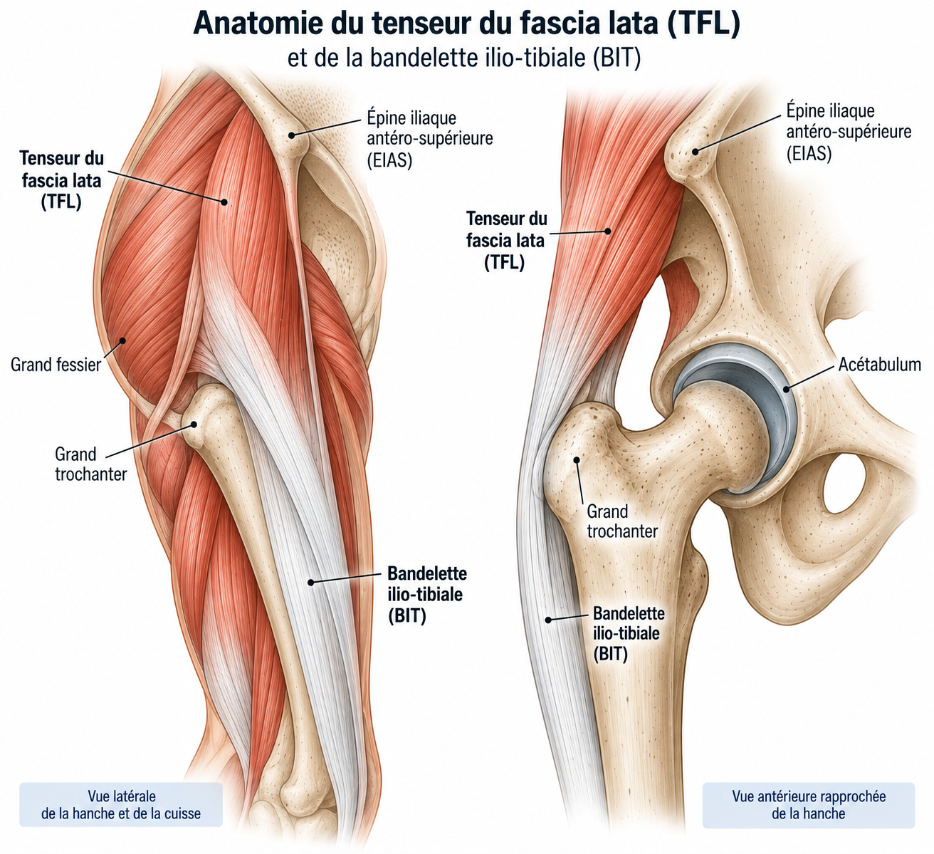
🔍 Click on the image to enlarge it
How does it differ from iliotibial band syndrome?
This distinction is important. Classic iliotibial band syndrome essentially refers to pain in the lateral compartment of the knee, at the lateral femoral epicondyle. TFL impingement instead corresponds to high, anterolateral pain near the ASIS, sometimes a few centimetres below the iliac crest — although the pain may nonetheless extend down the lateral thigh.
There is therefore an anatomical continuity between the two conditions: the TFL forms the proximal part of the musculofascial system that continues as the iliotibial band, but their clinical location is different.
Epidemiology
Anterolateral TFL impingement remains a little-studied entity: its true frequency is not precisely known, unlike other causes of lateral hip pain that are better characterised in the literature, such as greater trochanteric pain syndrome or iliotibial band syndrome at the knee.
It is mainly described in runners, particularly when the TFL is repeatedly loaded in movements combining hip flexion, abduction and internal rotation — such as during accelerations, sprints or hill running — or when its role in stabilising the pelvis becomes excessive because of a deficit in proximal control.
Musculotendinous or myofascial involvement of the TFL remains a less common cause of anterolateral hip pain than gluteus medius conditions or certain intra-articular conditions, which partly explains why it is often underdiagnosed or confused with other causes of lateral hip pain.
Causes & risk factors
Biomechanics in runners
The TFL is not usually “too short” or “too tight” in isolation: the problem is more one of excessive loading relative to the capacity of the gluteal system and the lower limb to absorb and control load. Several situations can increase its workload.
Sudden increase in load
This is probably the most common factor: an increase in mileage, a return after a break, more frequent sessions, the addition of interval sessions, a return to trail running, a large increase in elevation gain or a change of terrain.
Hill running
Uphill running in particular increases the demand on hip flexion and can increase the load on the hip flexors and stabilisers.
Accelerations and sprinting
The TFL contributes to hip control during acceleration phases. Sessions that include sprints, fast hills, changes of pace, fartlek or speed work can therefore become particularly symptomatic.
Gluteus medius control deficit
The gluteus medius is a major stabiliser of the pelvis. When its recruitment or endurance is insufficient, the TFL may play a greater part in controlling the hip. However, the condition should not be reduced to simple “gluteus medius weakness”: running biomechanics are multifactorial, and studies on the biomechanical factors in iliotibial band syndrome have in fact sometimes shown contradictory results.
Training factors
A rapid increase in volume, a simultaneous increase in volume and intensity, a return that is too quick, more hill sessions, more interval sessions, a sudden change of terrain or fewer recovery days can promote the onset of symptoms.
Biomechanical factors
Poor hip abductor endurance, poor pelvic control, excessive adduction of the lower limb during stance, excessive dynamic internal rotation, a running strategy that places heavy demands on the lateral stabilisers or significant muscle fatigue may also play a role.
Morphological factors
Certain individual characteristics can alter hip mechanics and the tension placed on the fascia lata. However, there is no single “TFL runner” body type.
Symptoms
The clinical presentation is usually fairly characteristic.
Location
The pain is located at the anterolateral part of the hip, near the ASIS, slightly behind or below it, and sometimes at the upper part of the lateral thigh. The patient can sometimes point to the painful area with one finger: this very precise location is a useful diagnostic clue.
Type of pain
The pain may be mechanical, localised, burning, tension-like, sometimes stabbing, and sometimes accompanied by a “pulling” sensation. It may spread towards the lateral thigh.
Triggering activities
Symptoms may appear during running, accelerations, uphill running, stairs, brisk walking, standing up from sitting, certain prolonged positions or hip abduction exercises. In some runners, the pain only appears after several kilometres.
Clinical examination
The examination should aim to reproduce the pain and, above all, to determine which structure is actually responsible.
Palpation. This looks for tenderness at the origin of the TFL, in front of the greater trochanter, along the proximal part of the iliotibial band or over the fascia lata. Very localised tenderness over the TFL is a pointer towards the diagnosis.
Resisted contraction. The TFL can be tested by combining hip flexion, abduction and internal rotation. Reproduction of pain during a resisted contraction suggests musculotendinous involvement. Hip flexion, abduction and internal rotation can also be tested separately, to determine whether the pain is genuinely related to TFL recruitment.
Assessment of the gluteus medius and pelvic control
The examination should not be limited to the TFL. The clinician should look for a Trendelenburg sign, loss of single-leg stability, femoral adduction, dynamic internal rotation, contralateral pelvic drop or trunk compensation. Useful functional tests include single-leg stance, the single-leg squat, the step-down, the step-up, the lunge and the single-leg hop. The aim is not only to measure maximal strength: endurance and motor control are often more important in runners.
Running analysis
Running analysis is particularly useful. It can look for excessive hip adduction, dynamic internal rotation, insufficient pelvic control, an excessive increase or reduction in step width, crossover, and changes that appear with fatigue. It is useful to observe the runner at the start of the run, after several minutes, and possibly after symptoms appear: fatigue can reveal control deficits that are not visible at rest. Running analysis is also one of the strongly recommended elements in the recent French consensus on the rehabilitation of iliotibial band conditions.
Further tests
Indications for imaging
In simple, typical cases, imaging is not always necessary. It becomes useful when the diagnosis is uncertain, when symptoms persist despite well-conducted rehabilitation, when the pain is severe, when the presentation is atypical, or when an intra-articular condition is suspected.
Ultrasound
Particularly useful: it allows assessment of the TFL, its origin, the fascia lata, the proximal part of the iliotibial band and the subcutaneous tissues, possibly dynamically during movement, and comparison of the painful side with the opposite side. It is particularly useful when superficial or proximal involvement of the TFL–fascia lata system is suspected.
MRI
May show muscle oedema, enthesopathy, abnormalities of the fascia lata, involvement of the proximal iliotibial band, muscle injuries, a gluteus medius condition, bursopathy or an associated intra-articular condition. Proximal involvement of the iliotibial band or its origin may in particular show up as oedema around the gluteal fascia and the origin of the TFL.
Differential diagnoses
Classic iliotibial band syndrome
Pain in the lateral compartment of the knee, at the lateral femoral epicondyle — TFL impingement, by contrast, corresponds to high, anterolateral pain near the ASIS, sometimes a few centimetres below the iliac crest.
Intra-articular hip conditions
Femoroacetabular impingement, labral tear, early osteoarthritis, synovitis or cartilage injury — to be considered in particular in the presence of deep groin pain, marked restriction of hip mobility or night pain.
Peri-articular hip tendinopathies
Tendinopathy of the gluteus medius, gluteus minimus, rectus femoris, iliopsoas or adductors, to be differentiated by the precise location of the pain and specific resisted contraction tests.
Other diagnoses to consider
Greater trochanteric pain syndrome, snapping hip, athletic groin pain (pubalgia), femoral neck stress fracture, pelvic bone stress injury, referred lumbar pain or meralgia paraesthetica.
Classification
Grade 1 – Mild irritation
Pain only after running, no pain in daily life, little tenderness on examination, strength preserved.
Grade 2 – Functional impairment
Pain during running, reduced distance covered, pain during certain daily activities, pain on resisted contraction, impaired dynamic control.
Grade 3 – Significant impairment
Pain appearing quickly on exertion, marked limitation of running, persistent pain after exercise, marked functional deficit, suspected associated tendon or muscle injury.
Treatment
Phase 1 – Reducing irritation
Treatment is based mainly on a temporary reduction in mechanical stress, followed by a gradual restoration of the capacity of the hip–pelvis system. The aim is not to "permanently relax the TFL", but to make the system able to tolerate the stresses of running again.
For a few days to a few weeks depending on severity, interval training, sprinting, hills, fast descents, technical trail running, very long sessions and exercises that clearly reproduce the pain should be temporarily reduced. Complete rest is rarely necessary: pain-free cardiovascular activity can be maintained (cycling if pain-free, swimming, elliptical trainer, walking), following the principle of relative rest. This approach is also recommended in the recent French consensus on iliotibial band conditions.
Phase 2 – Restoring mobility
Any restriction in mobility of the hip, the anterior chain, the quadriceps, the lateral fascia or the ankle should be looked for. Mobility exercises can be offered: gentle TFL stretching, hip flexor stretching, active hip mobility and hip rotation mobilisation.
Be careful with aggressive stretching: the TFL is a structure that can become painful when it is strongly compressed or put under tension. Stretching should therefore remain gradual and pain-free, without any feeling of anterolateral pinching.
Phase 3 – Targeted strengthening
Strengthening should gradually restore the capacity of the TFL, but above all that of the muscles involved in hip control. Simple exercises can be offered without significant pain: side-lying hip abduction (3 sets of 10 to 15 repetitions), clamshell (3 × 12-15), glute bridge (3 × 12-15), banded abduction (3 × 10-15).
A common question is whether the TFL should be strengthened or stretched: both can have a place, but the problem is not usually a TFL that is simply "too strong" or "too short". A painful TFL may be overloaded, lacking in endurance, subjected to excessive load, or used to compensate for a deficit in proximal control. Treatment must therefore address the whole chain: it is often more relevant to talk about functional rebalancing than about "deactivating the TFL".
Phase 4 – Functional strengthening and gluteus medius work
Once the simple exercises are well tolerated, the load is gradually increased with exercises such as the squat, single-leg squat, step-up, step-down, lunges, lateral lunges, stair climbing or single-leg deadlift. The aim is to work on strength, endurance, pelvic stability, knee control and trunk control at the same time.
The gluteus medius plays a central role in frontal-plane control of the pelvis, and its strengthening is progressed in levels: side-lying abduction and clamshell first, then standing banded abduction, monster walk and crab walk, then single-leg squat, step-down, lunge and single-leg deadlift, and finally single-leg hops, lateral bounds and plyometric work once the previous levels have been mastered.
In some runners, the problem appears mainly with fatigue: endurance should then be trained with long sets, single-leg exercises, pelvic holding, trunk control and functional repetitions, so that the runner can maintain good mechanics after several minutes of running, and not just during the first repetitions of an exercise.
Phase 5 – Running-specific work
The work should then move closer to the demands of running: double-leg jumps, single-leg hops, bounds, stride work, progressive accelerations and changes of direction can be introduced. Progression should go from simple to complex and from slow to fast — a progression in exercise complexity that is one of the recommendations to reach strong agreement in the 2025 French Delphi consensus.
Stride analysis can sometimes help to identify an aggravating mechanism. Depending on the case, a slight increase in cadence, a temporary reduction in stride length, less crossover, improved pelvic control or a change in foot path can be tried. These changes must be individualised: there is no universal running technique that prevents TFL pain.
Gradual return to running
The return should be gradual and change only one variable at a time: first increase duration, then distance, then frequency, then intensity. An example progression: alternating 2 minutes of running and 1 minute of walking for 20 to 30 minutes (step 1), then 5 minutes of running to 1 minute of walking (step 2), continuous low-intensity running (step 3), a gradual increase in duration (step 4) and then in weekly volume (step 5), reintroduction of hills (step 6), then of interval training (step 7), before returning to specific sessions (step 8).
Before returning to normal running, the following should be checked: no pain in daily life, little or no tenderness on palpation, a pain-free or almost pain-free resisted contraction, good abductor strength, good pelvic endurance, a controlled single-leg squat, a correctly performed step-down, tolerated single-leg hops, and easy running without a gradual increase in pain. Being able to perform the rehabilitation exercises without pain is not enough: the specific demands of running must also be tested.
Complementary treatments
Ice
May temporarily reduce pain after exercise, but does not treat the mechanical cause.
Anti-inflammatories
May be used occasionally during painful phases when appropriate, but should not replace load modification and rehabilitation.
Massage
Massage of the TFL or the surrounding tissues may provide symptomatic relief, but should not be considered the main treatment. Aggressive deep friction techniques on the iliotibial band, in particular, are debated and are not a central rehabilitation strategy.
Shockwave therapy
Their value in this specific indication is not well established. They may possibly be considered in certain chronic tendinopathies, but there is not enough evidence to make them a routine treatment for anterolateral TFL impingement.
Ultrasound-guided injection
May exceptionally be considered when the diagnosis is uncertain or when a specific structure is identified as the source of pain. It should remain an adjunct and not replace correction of the mechanical overload. Interventional ultrasound makes it possible to confirm the location of the abnormality, assess the TFL dynamically, compare both sides and guide a diagnostic or therapeutic injection, thus avoiding treating "blindly" lateral hip pain that may in fact come from the gluteus medius, the hip joint or another structure.
Surgery
Surgery is exceptional in anterolateral TFL impingement. It should only be considered after diagnostic confirmation, exclusion of differential diagnoses, properly conducted rehabilitation, load adaptation and prolonged failure of conservative treatment.
There is no standardised surgical treatment specifically established for this entity: any intervention would depend on the anatomical lesion identified.
Recovery time
The prognosis is generally favourable when the overload mechanism is identified. Recovery time depends in particular on how long the symptoms have been present, the level of sport, training volume, the presence of a tendon injury, any strength or endurance deficit, and the presence of an associated condition.
Recent pain related to overload generally responds better to early load adaptation. Conversely, chronic forms often require longer and more gradual rehabilitation.
Prevention
Prevention relies mainly on improving the system’s capacity to tolerate load: a gradual increase in mileage, avoiding simultaneous increases in volume and intensity, keeping recovery days, regularly strengthening the gluteal muscles and training the endurance of the hip stabilisers.
It is also recommended to introduce hills gradually, not to go straight back to fast sessions after a break, and to watch for the onset of reproducible pain. Prevention does not mean systematically stretching the TFL before every run.
Frequently asked questions
Is TFL impingement the same as iliotibial band syndrome?
Can the TFL cause hip pain?
Do I need to stop running completely?
Can I keep running with mild pain?
Is the TFL necessarily too tight?
Is gluteus medius strengthening always necessary?
Is an MRI mandatory?
How long does it take to return to running?
Common mistakes
Stopping running completely for several weeks: rest may settle the pain but does not necessarily restore the system's capacity.
Going straight back to the previous level: the disappearance of pain does not mean that load capacity has returned to its initial level.
Massaging the TFL very hard: an irritated structure does not necessarily need to be aggressively "unstuck".
Focusing solely on the TFL, without examining the gluteus medius, gluteus maximus, pelvic control, trunk, knee, ankle and running technique.
Systematically correcting the stride: a biomechanical change is only useful if it matches the runner's individual mechanism.
Treating the imaging alone: an abnormality of the TFL or fascia lata on MRI is only relevant if it matches the clinical picture.
Key points — Institut Basque du Coureur
Anterolateral impingement of the tensor fasciae latae is a possible cause of pain in the anterolateral part of the hip in runners. It generally corresponds to overload of the TFL–fascia lata system, rather than a simple abnormality of the muscle's length or tension. Diagnosis is based mainly on the very precise location of the pain, reproduction of symptoms when the TFL is loaded, examination of pelvic and hip control, running analysis, and exclusion of intra-articular conditions and other causes of lateral pain. Treatment is based above all on temporary load adaptation, reduction of triggering stresses, strengthening of the hip abductors and stabilisers, endurance and pelvic control work, functional progression and a gradual return to running. The aim is not to relax the TFL at all costs, but to make the whole hip–pelvis–lower limb unit strong, enduring and coordinated enough to tolerate the stresses of running again.
- Guyot A, et al. La rééducation du syndrome de la bandelette ilio-tibiale : recommandations formalisées par la méthode Delphi. J Traumatol Sport. 2025.
- Baker RL, Fredericson M. Iliotibial Band Syndrome in Runners: Biomechanical Implications and Exercise Interventions. Phys Med Rehabil Clin N Am. 2016;27(1):53-77.
- Fredericson M, Weir A. Practical management of iliotibial band friction syndrome in runners. Clin J Sport Med. 2006;16(3):261-268.
- Long S, Leahy H, Bush C, Surrey D, Nazarian L. Anterolateral hip pain: Sonographic evaluation of the proximal iliotibial band and tensor fascia lata. J Clin Ultrasound. 2020;48(4):193-197.
- Decker G, Hunt D. Proximal Iliotibial Band Syndrome in a Runner: A Case Report. PM R. 2019;11(2):206-209.
- Willy RW, et al. Current Clinical Concepts: Synthesizing the Available Evidence for Improved Clinical Outcomes in Iliotibial Band Impingement Syndrome. J Athl Train. 2021;56(8):805-815.
- Beyer R, et al. Beyond Greater Trochanteric Pain Syndrome: The Role of Tensor Fascia Latae Myofascial Pain in Lateral Hip Pain. Turk J Osteoporos. 2024.