
Definition
Gluteus medius tendinopathy is a painful condition of the tendon of the gluteus medius muscle, most often at its insertion on the greater trochanter. It is often part of a broader clinical picture called greater trochanteric pain syndrome (Greater Trochanteric Pain Syndrome, GTPS), which may involve the gluteus medius tendon, the gluteus minimus tendon, both tendons, the trochanteric bursa and the surrounding tissues.
The term “tendinitis” is now considered less precise: in chronic forms, the condition corresponds more to a change in the structure and mechanical capacity of the tendon than to simple inflammation. The term gluteal tendinopathy is therefore preferred; it is one of the main causes of lateral hip pain.
Anatomy
The gluteus medius is a muscle located on the lateral side of the pelvis. It originates from the outer surface of the ilium and inserts mainly on the lateral surface of the greater trochanter, an area under particularly high stress during single-leg stance. It has several bundles of fibres whose orientation allows different functions, and it works in synergy with the gluteus minimus, the gluteus maximus and the deep hip rotators.
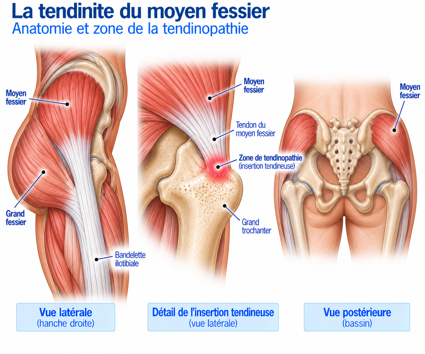
🔍 Click on the image to enlarge it
Function of the gluteus medius
The gluteus medius plays several essential roles. It produces hip abduction, i.e. moving the thigh away from the midline of the body. When a person is standing on one leg, it also ensures stabilisation of the pelvis by preventing it from tilting towards the opposite side.
Finally, it contributes to control of the lower limb: hip adduction, rotation of the femur and dynamic alignment of the knee. This function is particularly important when walking, running, climbing stairs, jumping and changing direction.
The gluteus medius in runners
When running, each foot strike places significant stress on the pelvis and hip. The gluteus medius must in particular stabilise the pelvis, control femoral movement and contribute to force transmission between the trunk and the lower limb; its activity increases with running speed.
However, it is important not to reduce running injuries to a simple “weak gluteus medius”: the available data do not show a universal, consistent deficit in activation of this muscle across the various running-related injuries.
Epidemiology
Gluteal tendinopathy is particularly common in middle-aged and older adults, and much more common in women: some publications report that it may affect up to one in four women over the age of 50, making it one of the most common tendinopathies of the lower limb.
However, it is by no means limited to postmenopausal women: it also affects men, athletes, runners, young people and people who do little sport.
Causes & risk factors
The gluteal tendon has to withstand tensile, compressive and shear stresses. Compression can be particularly high when the hip is in adduction, in flexion-adduction or in certain crossed positions — a classic example being sleeping for long periods on the painful side, which places the hip in adduction and compresses the tendon against the greater trochanter.
The problem can be summarised as follows: excessive or poorly tolerated tendon load + repeated compression → reduced capacity of the tendon to withstand stress → pain.
Compression: a key concept
Some seemingly harmless positions can strongly compress the gluteal tendons: crossing your legs, sleeping on the painful side, standing with your weight “hanging” on one hip, standing for long periods with the pelvis shifted to one side, some stretches in adduction or certain sitting positions. The first step of treatment therefore often consists of temporarily reducing these compressive stresses.
Load-related factors
Increased mileage, increased intensity, returning after a break, significant elevation gain, hill running, an increased number of sessions.
Mechanical factors
Marked hip adduction, prolonged single-leg stance, poor tolerance of certain positions, a sudden increase in lateral load.
Individual factors
The following are found more frequently: middle or older age, female sex, a history of low back pain, knee or hip osteoarthritis, and excess body weight in some populations. However, these epidemiological associations remain complex and should not be interpreted as direct causes.
Symptoms
Patients often say: “My hip hurts on the side”, then “when I sleep on that side, it wakes me up”, or “it hurts when I go up stairs or when I run”. In runners, the typical scenario combines an increase in training volume, lateral hip pain, pain during and then after running, and discomfort on stairs or when lying down.
The pain is usually located on the outer side of the hip, around the greater trochanter. It can sometimes radiate down the lateral side of the thigh to the knee, or be felt further back, in the buttock region. It is described as dull, burning, throbbing, deep or, on the contrary, very localised.
Night pain
Night pain is highly characteristic: the patient may feel pain when sleeping on the painful side, when turning over in bed, or sometimes even when sleeping on the opposite side — because the painful leg can then drop into adduction and compress the tendon against the greater trochanter. A pillow between the knees can sometimes reduce this compression.
Pain when walking and running
Single-leg weight-bearing activities place heavy demands on the hip abductors: symptoms may therefore appear during prolonged walking, running, on stairs, going uphill or downhill, or when changing direction. In runners, pain may appear during the run, after the run, several hours later, or sometimes only the following morning.
Clinical examination
The examination starts by determining the exact location of the pain and looking for night pain, pain when walking, running, standing on one leg or using stairs, as well as a history of lumbar problems or intra-articular hip symptoms. Hip mobility, abductor and rotator strength, and control of the pelvis and lower limb are then examined.
Palpation of the greater trochanter is a very important element: pain that precisely reproduces the usual pain greatly increases the likelihood of gluteal involvement, while the absence of tenderness on palpation makes the diagnosis less likely.
The single-leg stance test (about 30 seconds) looks for the onset of pain, pelvic stability, hip adduction and any trunk compensation; reproduction of pain within the first few seconds is highly suggestive. Resisted hip abduction looks for pain, weakness and asymmetry. The combination of pain on palpation of the greater trochanter + pain on resisted abduction is particularly useful for confirming the diagnosis clinically.
The examination can be completed with functional tests: single-leg squat (pain, pelvic control, hip adduction, knee stability), step-down (frontal-plane control of the lower limb), walking (looking for a limp) and running (pelvis, cadence, step width, foot-contact control, onset of pain).
A drop of the pelvis on the opposite side may reflect abductor insufficiency, but does not automatically mean that “the gluteus medius is weak and simply needs strengthening”: strength, pain, coordination, fatigue and individual strategies interact, which calls for individualised management.
Further tests
Ultrasound
Looks for tendon thickening, changes in echotexture, hypervascularity, a tear, a rupture or associated bursitis. Also allows dynamic assessment.
MRI
Assesses the gluteus medius, gluteus minimus, the bursae, muscle quality and any tear. Particularly important when a partial or complete tear is suspected or surgery is being considered. Note: an MRI abnormality is not necessarily responsible for the pain, as such abnormalities can also be found in people without symptoms.
Differential diagnoses
Gluteus minimus tendinopathy
Very often associated with gluteus medius tendinopathy; the two frequently coexist in the same clinical picture.
Trochanteric bursitis
May coexist with tendinopathy but is rarely the sole explanation: in a large ultrasound series, fewer than a quarter of patients with greater trochanteric pain syndrome had isolated bursitis, gluteal tendon abnormalities being much more common.
Iliotibial band syndrome
Can cause lateral hip pain, particularly during certain dynamic movements.
Hip osteoarthritis
To be considered in the presence of groin pain, limited rotation, stiffness or deep pain.
Femoroacetabular impingement
Can cause hip pain that is sometimes referred to the side.
Labral tear
To be considered in the presence of deep pain, a click, catching or pain during rotational movements.
Referred lumbar pain / radiculopathy
A lumbar origin can mimic hip pain; look for radiating pain, paraesthesia or a neurological deficit.
Gluteal tendon tear or rupture
To be suspected in the presence of marked weakness in abduction, a limp, a Trendelenburg sign, difficulty standing on one leg or muscle atrophy — a different situation from simple tendinopathy.
Classification
Stage 1 — Mild irritation
Pain after activity, little discomfort in daily life, sleep barely disturbed.
Stage 2 — Moderate tendinopathy
Pain during certain activities, discomfort when running, night pain, difficulty with stairs.
Stage 3 — Significant tendinopathy
Daily pain, significant limitation of walking, disturbed sleep, functional impairment.
Stage 4 — Significant structural damage
Partial or complete tear, marked weakness, Trendelenburg sign, major functional limitation.
Treatment
Reducing compression
This step is often underestimated, even though it is the first key to management. The following should be temporarily limited: sleeping on the painful side, crossing your legs, standing "slumped" on one hip, certain prolonged positions in adduction and certain aggressive gluteal stretches.
For sleeping, you can use a pillow between your knees, and possibly a pillow behind your back to avoid rolling onto the painful side. The aim is to reduce compression without stopping all activity.
Adapting running rather than stopping
In runners, distance, speed, elevation gain, session frequency, terrain and pain during and after exercise need to be reassessed. A temporary reduction in volume may be enough — for example, going from "a painful 10 km" to "a tolerated 5–6 km" rather than stopping completely for several weeks.
Yes, in many cases you can run with gluteal tendinopathy: mild, controlled pain can be compatible with adapted activity. The main things to monitor are how the pain changes during the session, pain in the following hours, night pain and how you feel the next morning. A gradual worsening or an increase in night pain generally means that the load is still too high.
Pain relief and muscle activation
When the tendon is very irritable, treatment starts with isometric contractions: isometric abduction (pushing the knee against resistance without movement, 4–5 × 30–45 seconds), bridges (3 × 10–15) or abduction in a controlled position with light resistance. The initial aim is to regain a tolerable muscle contraction without significantly increasing symptoms.
Progressive strengthening
Resistance is then gradually increased: hip abduction with a resistance band, side steps, hip thrusts, single-leg bridges, weight-bearing abduction, step-ups, squats, lunges. The aim is to turn a muscle capable of producing moderate force into a system capable of withstanding the stresses of sport.
Single-leg work and the whole kinetic chain
This phase is particularly important: single-leg stance, single-leg squats, step-downs, step-ups, lunges, pelvic control — aiming for stability, knee control, pelvic control and no significant pain.
The gluteus medius should not be rehabilitated in isolation: hip work (gluteus medius, gluteus maximus, rotators) is combined with knee work (quadriceps, hamstrings), ankle work (triceps surae, foot control) and trunk work (pelvic control, lumbopelvic stability), in order to improve force transmission throughout the whole chain.
Dynamic work and return to running
Once strength is sufficient, jumps, bounds, changes of direction, accelerations and decelerations are introduced; for runners: strides, hills and interval training. The tendon must gradually be exposed to the stresses required for a return to sport.
The return to running follows a progression: brisk walking without worsening, alternating walking and running, slow continuous running, increasing duration and then weekly volume, introducing speed, hills, interval training, and then returning to specific sessions.
Before resuming full training, the aim is to have mild, stable pain with no worsening the next day, good abduction strength and single-leg stance, the ability to perform single-leg squats, step-downs, lunges, jumps and landings, and the ability to run progressively without a lasting increase in symptoms.
Complementary treatments
Manual therapy
Can be used as a complement to temporarily reduce pain, improve mobility and make exercises easier, but should not replace strengthening work.
Taping
Can temporarily change pain, the feeling of stability or tolerance of certain activities, particularly at the start of rehabilitation. It is not a long-term treatment.
Extracorporeal shockwave therapy
A worthwhile option in persistent forms, after failure of properly conducted management. The data are encouraging but inconsistent; a recent systematic review suggests a long-term benefit from some forms of focused shockwaves, but comparisons between techniques remain difficult.
Corticosteroid injection
Can provide relatively rapid relief, which is useful in cases of very severe pain, difficulty sleeping or inability to carry out rehabilitation. However, its effect is often temporary: in the randomised trial by Mellor et al., it was more effective than a wait-and-see approach in the short term, but the education + exercise programme gave better overall results at 8 and 52 weeks. It should not replace active treatment.
PRP (platelet-rich plasma)
Some studies suggest a benefit, particularly in the medium or long term, but protocols remain highly variable. A 2025 systematic review considers it promising, particularly compared with corticosteroids on certain short-term functional outcomes, but highlights the need for more robust trials. It can be discussed in certain resistant chronic forms, without being presented as an essential treatment.
Surgery
Surgery is rarely needed for simple gluteal tendinopathy. It may be considered in cases of a large partial tear or a complete tear, major functional weakness, prolonged failure of conservative treatment or significant limitation of daily life or sport. Possible techniques include endoscopic repair, open repair, debridement, tendon reattachment or augmentation for some large tears — although the surgical data remain much less robust than those for conservative treatment.
Certain characteristics can complicate recovery: long duration, a large tear, retraction, muscle atrophy, fatty infiltration, marked weakness or late diagnosis — the quality of the muscle and tendon being particularly important when surgical repair is being considered.
Rehabilitation after surgery is generally progressive: protecting the repair and reducing weight-bearing according to the surgeon’s instructions (phase 1), muscle contractions and trunk work (phase 2), strengthening of the abductors, gluteus maximus, quadriceps and posterior chain (phase 3), single-leg work — balance, step-ups, step-downs (phase 4), then reintroduction of impact — jumps, running, accelerations, sport-specific work (phase 5). The timeline depends heavily on the type of injury and the surgical technique.
Recovery time
Improvement may be seen within a few weeks, but chronic forms often require several months to regain full capacity. The return to running should never be guided solely by the disappearance of pain, but by the tissues’ progressive ability to tolerate the stresses of daily life and sport.
Prevention
Prevention is based mainly on load management. Runners should avoid increasing mileage, speed, elevation gain and session frequency at the same time.
It is also helpful to maintain hip strengthening, general strength, regular single-leg work and sufficient recovery.
Night pain, pain in daily life and pain after sessions should be monitored particularly closely: night pain in particular is an excellent indicator of tendon irritability.
Frequently asked questions
Is it really a tendinopathy?
Is it bursitis?
Why does it hurt when I sleep on my side?
Why does it hurt when I cross my legs?
Should I stop running?
Is strengthening the gluteus medius enough?
Is stretching helpful?
Can an injection heal the tendon?
Does PRP work?
How long does it take to heal?
Can a tear occur without trauma?
Common mistakes
Thinking it is simple bursitis, when gluteal tendinopathy is very often the main problem.
Massaging the painful tendon directly: repeated pressure on the greater trochanter can increase compression.
Sleeping on the painful side, one of the most classic sources of compressive stress.
Habitually crossing your legs, which increases adduction and tendon compression.
Doing a lot of stretching, when some stretches can increase compression of the tendon.
Doing only abduction exercises, when the tendon needs to gradually regain overall functional capacity.
Stopping moving altogether, which reduces muscle and tendon capacity.
Relying solely on MRI, when gluteal abnormalities can also be present in people without symptoms.
Having an injection and then immediately returning to maximum level: the temporary disappearance of pain does not mean that the tendon's capacity has been restored.
Key points — Institut Basque du Coureur
Gluteus medius tendinopathy, more broadly known as gluteal tendinopathy, is a very common cause of lateral hip pain, often wrongly presented as simple "trochanteric bursitis". Diagnosis is based mainly on lateral hip pain, tenderness on palpation of the greater trochanter, pain in single-leg stance, pain on resisted abduction and analysis of hip function. Imaging can confirm tendon involvement or look for a tear, but an MRI abnormality is not necessarily responsible for the symptoms. Treatment is based above all on reducing compression, load management, progressive strengthening and functional return: the aim is not to protect the tendon indefinitely, but to gradually restore its capacity to withstand the stresses of daily life and sport. In the presence of marked weakness, a limp or a Trendelenburg sign, a gluteal tendon tear or rupture should be looked for, which is a different situation from simple tendinopathy.
- Grimaldi A, Fearon A. Gluteal Tendinopathy: Integrating Pathomechanics and Clinical Features in Its Management. J Orthop Sports Phys Ther. 2015;45(11):910-922.
- Grimaldi A, Mellor R, Hodges P, Bennell K, Wajswelner H, Vicenzino B. Gluteal Tendinopathy: A Review of Mechanisms, Assessment and Management. Sports Med. 2015;45(8):1107-1119.
- Mellor R, Bennell K, Grimaldi A, et al. Education plus exercise versus corticosteroid injection use versus a wait and see approach on global outcome and pain from gluteal tendinopathy. Br J Sports Med. 2018;52:1464-1472.
- Cordeiro TTP, Rocha EAB, Silva RS. Effects of exercise-based interventions on gluteal tendinopathy: systematic review with meta-analysis. Sci Rep. 2024;14:3343.
- Dancy ME, Alexander AS, Clark CJ, et al. Gluteal Tendinopathy: Critical Analysis Review of Current Nonoperative Treatments. JBJS Rev. 2023;11(10):e23.00101.
- Ladurner A, Fitzpatrick J, O'Donnell JM. Treatment of Gluteal Tendinopathy: A Systematic Review and Stage-Adjusted Treatment Recommendation. Orthop J Sports Med. 2021;9.
- Grimaldi A, Mellor R, Hodges P, et al. Utility of clinical tests to diagnose MRI-confirmed gluteal tendinopathy in patients presenting with lateral hip pain. Br J Sports Med. 2017.
- Fearon AM, Scarvell JM, Neeman T, Cook JL, Cormick W, Smith PN. Greater trochanteric pain syndrome: defining the clinical syndrome. Br J Sports Med. 2013.
- Westacott DJ, Minns JI, Foguet P. The diagnostic accuracy of magnetic resonance imaging and ultrasonography in gluteal tendon tears: a systematic review. Skeletal Radiol. 2011.
- Hilligsøe M, Rathleff MS, Olesen JL. Ultrasound Definitions and Findings in Greater Trochanteric Pain Syndrome: A Systematic Review. J Ultrasound Med. 2020;46:1584-1598.
- Semciw A, Neate R, Pizzari T. Running related gluteus medius function in health and injury: a systematic review with meta-analysis. J Electromyogr Kinesiol. 2016;30:98-110.
- The efficacy of gluteal tendinopathy treatments: A systematic review. Clin Rehabil. 2025;39(5):600-617.
- Fusco S, Idoate Saralegui F, Capurro-Soler B, Fiz N, Llopis E. Gluteal Tendons Tears: Imaging Diagnosis with Treatment Implications. Magn Reson Imaging Clin N Am. 2026.
Physiotherapy protocol for this injury
Découvrez notre protocole de rééducation détaillé pour gluteus medius tendinopathy : exercices adaptés, progression structurée et modalités de reprise.