
Definition
Femoroacetabular impingement, or FAI syndrome (FemoroAcetabular Impingement syndrome), refers to a situation in which the shape of the hip joint promotes abnormal contact between the femur and the rim of the acetabulum during certain movements, particularly those combining hip flexion, adduction and internal rotation. This contact can cause hip or groin pain, restricted mobility, a labral tear, cartilage damage and reduced sports performance.
It is essential to distinguish femoroacetabular morphology from femoroacetabular impingement syndrome itself. A cam or pincer morphology can perfectly well be present in someone who has no symptoms at all. A diagnosis of FAIS therefore requires three elements together: compatible symptoms, suggestive clinical signs and compatible morphological abnormalities on imaging. This definition is now the central principle of the international Warwick consensus on the subject.
Imaging alone does not make the diagnosis
An X-ray can show femoroacetabular impingement in a patient who has no pain at all: cam and pincer morphologies are common in people without symptoms, especially athletes. What matters is therefore not the image on its own, but the consistency between the clinical history, the examination, the observed morphology and the symptoms actually reported by the patient.
Anatomy
The hip is a ball-and-socket joint made up of the femoral head, femoral neck, acetabulum, labrum, articular cartilage, joint capsule and periarticular muscles. The acetabulum forms a socket that receives the femoral head; the acetabular labrum increases its depth and contributes both to joint stability and to its peripheral sealing effect. Cartilage covers the femoral head and the articular surface of the acetabulum. When mechanical contact between the femur and the acetabular rim becomes excessive or repetitive, it is precisely the labrum and the cartilage that are put under stress.
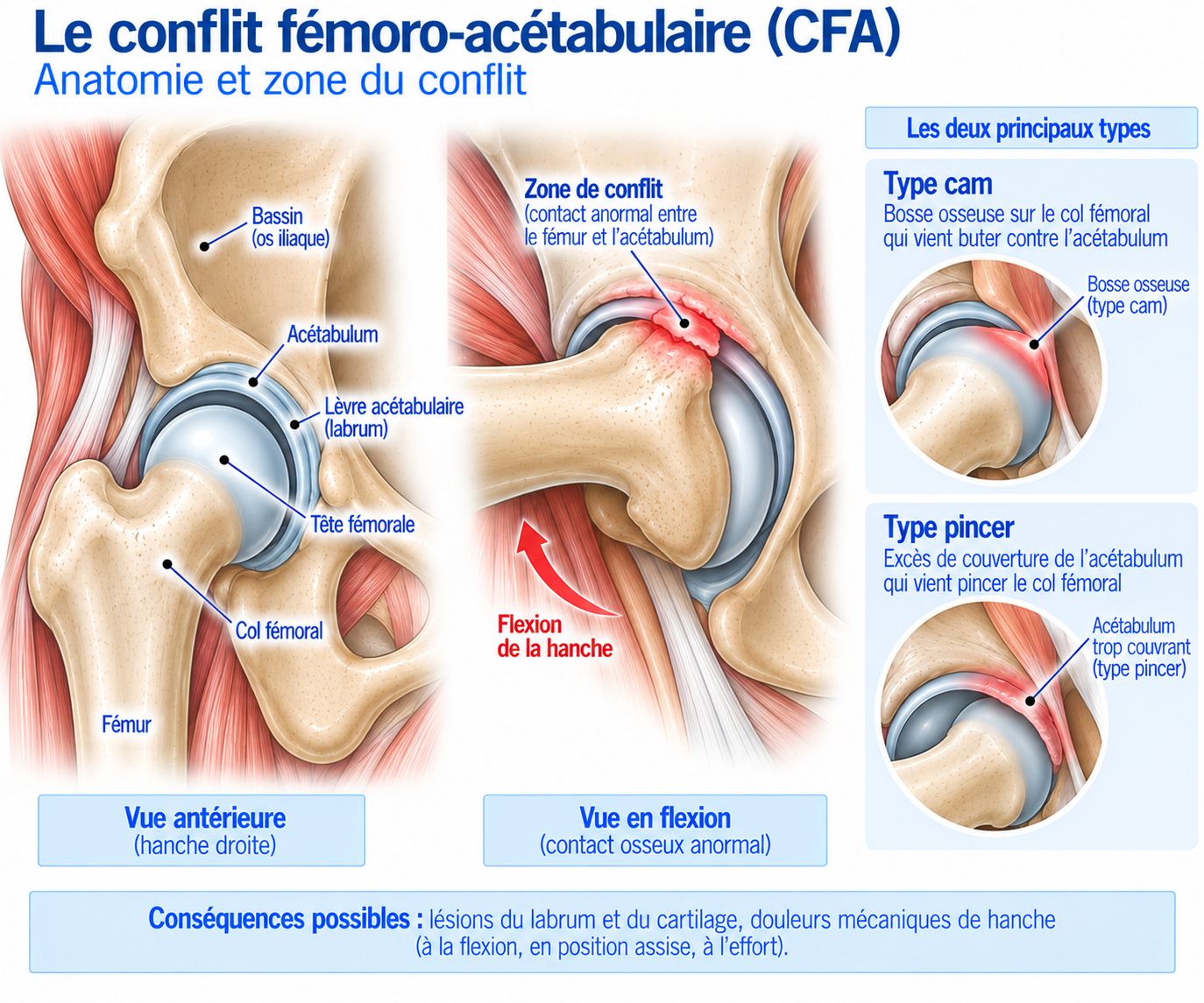
🔍 Click on the image to enlarge it
The three morphologies of femoroacetabular impingement
Three morphologies are traditionally recognised as causes of impingement.
Cam type — the term “cam” refers to an abnormality of the junction between the head and neck of the femur. Normally, the femoral head is broadly spherical; in cam morphology, this junction shows asphericity or a bony prominence. During flexion and internal rotation, this area can come into contact with the acetabular rim: the impingement is therefore mainly related to the shape of the femur.
Pincer type — this corresponds to acetabular over-coverage of the femoral head: the rim of the acetabulum can then come into early contact with the femoral neck. The mechanism is therefore mainly related to the shape of the acetabulum, which may result in particular from global over-coverage, acetabular retroversion, acetabular protrusion or certain anatomical variations of the pelvis.
Mixed type — this combines cam and pincer morphology. It is a common situation in populations with symptomatic FAIS.
Biomechanics of impingement
Impingement occurs mainly when the hip moves into a combination of movements that reduces the functional space available between the femur and the acetabulum: the classic position is flexion combined with adduction and internal rotation. This is why some patients trigger their pain when sitting low, leaning forward, putting on a sock, getting into a car, doing deep squats or performing certain sports movements.
In runners, the situation is more complex: running involves several thousand hip flexion-extension cycles per session, pelvic stabilisation, high joint loads, rapid muscle contractions and repeated impacts. Impingement can therefore become symptomatic when the cumulative load exceeds the system’s capacity to adapt. Symptoms can be aggravated by a sudden increase in mileage, speed work, hills, accelerations, a stride with marked hip flexion, strength training in deep flexion, deep squats or any exercise that places heavy demands on the hip in flexion-adduction. Bone shape does not change in a few weeks, however: what changes quickly is above all the tolerance to loading.
Epidemiology
FAIS is particularly common in young, active athletes. Cam and pincer morphologies are also common in athletes without symptoms: a systematic review and meta-analysis of young athletes published in 2026 found an estimated radiological prevalence of around 48% for cam morphology and 18% for pincer morphology. These figures relate to radiological morphologies and must on no account be equated with the prevalence of symptomatic FAIS.
Football, combat sports, hockey, dance and certain sports requiring a large hip range of motion are particularly affected. Running can also bring to light pre-existing FAIS, especially when training volume or intensity increases.
Causes & risk factors
Several factors can promote the onset of symptoms in a patient with a compatible femoroacetabular morphology.
Anatomical factors
Cam morphology, pincer morphology, mixed morphology, associated dysplasia, history of hip problems.
Sport-related factors
Intensive training, sports involving a large hip range of motion, a sudden increase in volume, a substantial increase in intensity, repeated work in deep flexion.
Functional factors
Weak gluteal muscles, poor pelvic control, poor lower-limb control, reduced hip mobility, poor tolerance of high loads.
Symptoms
The most typical symptom is pain at the front of the hip or in the groin. Patients often point to the painful area with one finger or describe a deep pain “in the crease of the groin”. The pain can also be anterolateral, in the buttock, or sometimes felt in the thigh.
It often appears during movements that bring the hip into marked flexion: prolonged sitting, driving, cycling, deep squats, deep lunges, getting into or out of a car, standing up from a low chair, or putting on shoes and socks. In runners, it can appear during accelerations, on uphill sections, after a long run or after a strength training session.
Some patients also describe locking or snapping sensations: clicking, catching, a feeling of locking, or the impression that the hip “catches”. These symptoms may be related to the labrum, a tendon structure, mechanical impingement or a snapping iliopsoas, so any hip clicking should not automatically be considered femoroacetabular impingement.
Clinical examination
The examination begins with an overall assessment: limping, pelvic posture, hip mobility, muscle strength, single-leg control, lumbar mobility, knee mobility, neurological signs.
In particular, flexion, extension, abduction, adduction, internal rotation and external rotation are measured. Reduced internal rotation, particularly in flexion, may point towards an intra-articular problem; however, restricted mobility is not specific to FAIS, as it may also be constitutional or related to other conditions.
The FADIR test (flexion, adduction, internal rotation) reproduces the classic impingement mechanism; a positive test may reproduce groin pain, anterior hip pain or restricted movement. It is useful for guiding the diagnosis but is not sufficient on its own, as it can be positive in various hip conditions. The FABER test (flexion, abduction, external rotation) can help to look for an intra-articular problem, restricted mobility, sacroiliac involvement or a periarticular condition; its interpretation depends in particular on where the reproduced pain is located.
The examination can be completed with the log roll test, a resisted flexion test, tests of the psoas, abductors, adductors and gluteus medius, and an assessment of single-leg control. The aim is not to find “the positive test”, but to build up a body of clinical evidence. As there is no universal clinical classification of FAIS, a functional approach using severity grades (see below) can help guide management.
Further tests
Pelvic X-rays
An anteroposterior pelvic X-ray combined with a view showing the femoral head-neck junction (Dunn view or an appropriate lateral view), recommended as the first-line investigation by the recent ISHA international consensus. It assesses the shape of the femoral head, the head-neck junction, acetabular coverage, and any osteoarthritis, dysplasia or retroversion.
Alpha angle and related measurements
The alpha angle assesses the asphericity of the femoral head-neck junction (a high angle is compatible with cam morphology); other measurements can complete the assessment: lateral center-edge angle (LCEA), anterior center-edge angle, acetabular roof (sourcil) angle, signs of retroversion, joint space. The threshold value depends on the measurement method, the view and the population studied: an isolated value should not be considered a diagnosis of disease.
MRI (± MR arthrography)
Looks for a labral tear, cartilage damage, bone oedema, a subchondral reaction, tendinopathy, a muscle problem or a stress fracture. MR arthrography improves visualisation of the labrum. However, a labral tear on MRI does not necessarily mean that it is responsible for the symptoms: labral abnormalities and impingement morphologies are regularly seen in people without symptoms.
Diagnostic intra-articular injection
In some complex situations, an intra-articular anaesthetic injection guided by ultrasound or X-ray can be used as a diagnostic test: clear relief of the usual pain during an activity that reproduces the symptoms supports the hypothesis of an intra-articular origin, which is useful when several structures could be responsible for the pain.
Differential diagnoses
Intra-articular conditions
Femoroacetabular impingement, labral tear, osteoarthritis, synovitis, cartilage damage, loose body: here, the differential diagnosis relies mainly on the consistency between imaging, examination and clinical history.
Musculotendinous conditions
Iliopsoas tendinopathy, adductor tendinopathy, rectus femoris injury, gluteus medius tendinopathy, muscle injury: often reproduced by resisted tests rather than by FADIR.
Bone conditions
Femoral neck stress fracture, pelvic stress fracture, bone oedema: these should always be considered in runners with a recent increase in load, and MRI then makes the difference.
Other causes of groin pain
Athletic pubalgia, hernia, sacroiliac pain, referred lumbar pain, ischiofemoral impingement, snapping hip: groin pain in runners can have many causes, which makes this step essential.
Classification
Grade 1 — Mild
Pain only during strenuous exertion, normal daily activity, relatively preserved mobility, sport still possible.
Grade 2 — Moderate
Pain during running, reduced training volume, pain when sitting for long periods, restriction of certain movements, reproducible clinical tests.
Grade 3 — Severe
Frequent pain, marked limitation of running, discomfort in daily life, clearly restricted range of motion, pain persisting after exertion.
Grade 4 — Advanced disease
Significant cartilage damage, associated osteoarthritis, major functional limitation, failure of properly conducted conservative management.
Treatment
Phase 1 — Reduce the irritating load
Treatment depends on many factors: age, sporting level, symptoms, morphology, labral and cartilage involvement, duration of symptoms and sporting goals. It may be conservative, rehabilitation-based or surgical; conservative management is generally the first step, particularly when there is no advanced damage.
The first step is to temporarily reduce the most provocative movements. In runners, this may mean cutting back on interval training, sprinting, hills, long runs, significant elevation gain, deep squat sessions and exercises involving marked hip flexion. It is generally not necessary to stop all activity: the aim is to maintain a level of physical activity that the hip can tolerate.
Running can often be continued if symptoms remain mild and stable, by temporarily reducing mileage, running more slowly, cutting out hills and accelerations, adding recovery days and favouring short runs. Pain that gradually increases during the session or persists clearly afterwards indicates that the load is probably still too high.
Phase 2 — Mobility work
Contrary to popular belief, the aim is not necessarily to regain as much range of motion as possible. In a patient with cam morphology, aggressively trying to increase deep flexion can sometimes reproduce the painful mechanism.
The emphasis is on comfortable mobility, active control, pain-free rotation work, and mobility of the ankle, spine and pelvis. The principle is to improve useful mobility without forcing into the impingement zone.
Phase 3 — Muscle strengthening
Strengthening is a central component of management. It targets in particular the abductors, hip extensors, rotators, trunk muscles, quadriceps and hamstrings.
Exercises may include: glute bridge (3 × 10–15), hip abduction (3 × 10–15), clamshell (3 × 12–15), squat with controlled range (3 × 8–12) and step-up (3 × 8–12 per side).
Phase 4 — Functional strengthening and lumbopelvic control
The progression then includes the single-leg squat, lunges, step-down, single-leg deadlift, step-ups and loaded exercises, with the aim of developing strength, endurance and control of the pelvis, knee and trunk. The ISHA physiotherapy recommendations place particular emphasis on strengthening, motor control and functional exercises rather than routinely seeking to increase joint range of motion.
The pelvis plays a major role in hip mechanics: the work focuses on single-leg stability, control of the pelvis and trunk, and anti-rotation and unilateral exercises, such as the side plank, Pallof press, single-leg squat, single-leg deadlift and step-down.
Phase 5 — Reintroducing impacts
Before a full return to running, two-footed jumps, single-leg hops, bounds, small lateral jumps, plyometrics and progressive accelerations are gradually reintroduced, to check that the hip tolerates rapid loading.
Phase 6 — Gradual return to running
The return can follow a progression such as: alternating 2 min running / 1 min walking for 20–30 minutes, then 5 min running / 1 min walking, then easy continuous running, then a gradual increase in duration, then in mileage, then reintroduction of hills, then accelerations, and finally intervals and sport-specific training. The principle is not to increase volume, intensity, elevation gain and frequency at the same time.
Before a full return, the ideal is to have no or almost no pain in daily life, satisfactory functional mobility, strength that is symmetrical or close enough to the opposite side, good single-leg control, a well-controlled single-leg squat, pain-free jumps, tolerated easy running and no delayed worsening of symptoms. For high-level athletes, the return must then include accelerations, decelerations, changes of direction, sport-specific movements, fatigue and the demands of the sport: return to sport must therefore be criteria-based and not based on time alone.
Complementary treatments
Painkillers / anti-inflammatories
Can be used occasionally when appropriate, to reduce pain during an irritable phase. However, they do not correct bone shape, motor control, muscle capacity or load management, and should not be used to keep up excessive training despite significant pain.
Intra-articular injection
May have a diagnostic, pain-relieving or sometimes short-term therapeutic purpose, and is particularly useful when there is uncertainty about the exact origin of the pain. It is not, however, a treatment for the cam or pincer morphology itself.
Surgery
Surgery for femoroacetabular impingement is mainly performed by hip arthroscopy. It can be used to correct cam morphology, treat over-coverage in certain situations, repair or preserve the labrum, treat certain cartilage lesions and treat certain associated conditions. The aim is to restore more favourable joint mechanics while preserving the structures of the hip as much as possible.
It may be considered when the symptoms are clearly attributable to FAIS, the morphology is compatible, the functional impairment is significant, structured rehabilitation has failed and the sporting or functional goals justify an intervention. It should not be offered simply because an X-ray shows cam or pincer morphology: the international consensus stresses the need for consistency between symptoms, examination and imaging.
Randomised trials show that both approaches, surgery and rehabilitation, can improve symptoms, but in certain selected symptomatic patients, arthroscopy gives better functional results in the short and medium term than physiotherapy alone: in the UK FASHIoN trial, arthroscopy was superior to a programme of physiotherapy and activity modification on the functional score at 8 months. This does not mean that all patients should have surgery: rehabilitation remains an important step for patients who respond to conservative treatment, to determine functional tolerance, to optimise strength before any surgery, and after the surgery itself. A meta-analysis of randomised trials also showed that physiotherapy improves symptoms and does not appear to compromise the results of later surgery in patients in whom conservative treatment fails.
Recovery time
Postoperative rehabilitation is gradual. The initial goals are to control pain, regain functional mobility, protect the repaired tissues and restore muscle control. The progression then includes mobility, muscle activation, strengthening, neuromuscular control, endurance, impacts, sport-specific exercises and finally return to sport; recently published ISHA recommendations propose a six-phase progression based both on time since surgery and on meeting functional criteria.
Timescales depend in particular on the type of procedure performed, any labral repair, the condition of the cartilage, associated procedures and the sport played. Return to daily activities comes well before return to sport; for high-impact sports or those requiring complex hip movements, full return may take several months and should not be decided solely on the basis of the pain having gone.
The outlook is generally good when the diagnosis is correct, the hip does not have osteoarthritis, cartilage damage is limited, management is started early and the patient follows a structured rehabilitation programme. It is less favourable in the presence of advanced osteoarthritis, significant loss of joint space, extensive cartilage damage, significant dysplasia, older age or complex chronic pain.
Prevention
Primary prevention of morphological FAIS is difficult, since bone shape cannot simply be “corrected” with exercises. On the other hand, it is probably possible to work on preventing symptoms, through gradual training progression, regular strengthening, good lumbopelvic control, good muscle capacity, varied loading, sufficient recovery, and by avoiding sudden increases in volume and intensity.
In adolescent athletes, load management during growth periods deserves particular attention: cam morphology is now thought to be able to develop, in particular during bone growth, in young people who take part in certain intensive sports.
Frequently asked questions
What is the difference between femoroacetabular impingement and cam morphology?
Is cam a malformation?
Can you have femoroacetabular impingement without pain?
Is femoroacetabular impingement responsible for all groin pain in runners?
Does femoroacetabular impingement inevitably lead to osteoarthritis?
Can you keep running with FAIS?
Should squats be avoided?
Should the hip be stretched?
Can cam morphology develop through sport?
Is surgery always necessary?
Can you return to running after surgery?
Can an injection cure the impingement?
Common mistakes
Confusing morphology with disease: a cam appearance on imaging does not mean that the patient has FAIS.
Operating on an X-ray rather than on a symptomatic patient.
Relying solely on FADIR, a test that can be positive in many hip conditions.
Trying at all costs to increase range of motion: forcing flexion or internal rotation can worsen symptoms in some patients.
Stopping all activity completely: complete rest does not restore muscle capacity.
Continuing to run despite pain that increases during exercise.
Neglecting the pelvic muscles: treatment is not only about the joint.
Treating all groin pain as FAIS, when the differential diagnosis is broad.
Key points — Institut Basque du Coureur
Femoroacetabular impingement is a common cause of anterior hip pain in athletes. There are three main morphologies: cam (abnormality of the femoral head-neck junction), pincer (acetabular over-coverage) and mixed (a combination of the two). But an abnormal morphology on imaging is not the same as disease: a diagnosis of true FAIS relies on consistency between compatible symptoms, a suggestive clinical examination and imaging showing a compatible morphology. In runners, first-line management relies on load adjustment, hip and pelvic strengthening, neuromuscular control, restoring functional mobility, a gradual return to impacts and a gradual reintroduction of running. Arthroscopic surgery can give better results in certain selected symptomatic patients when rehabilitation is not enough, but it should never be decided solely on the presence of cam or pincer morphology on an X-ray: the right goal is not to make the X-ray normal, but to make the hip strong, mobile and tolerant enough for the athlete to get back to their activity.
- Griffin DR, Dickenson EJ, O'Donnell J, et al. The Warwick Agreement on femoroacetabular impingement syndrome (FAI syndrome): an international consensus statement. Br J Sports Med. 2016;50(19):1169-1176.
- Martin M, Moore JAM, Gomes DA, et al. Prevalence of Cam and Pincer Morphology in the Young Athlete's Hip: A Systematic Review With Meta-Analysis. J Orthop Sports Phys Ther. 2026;56(6):346-358.
- Frank JM, Harris JD, Erickson BJ, et al. Prevalence of femoroacetabular impingement imaging findings in asymptomatic volunteers: a systematic review. Arthroscopy. 2015;31(6):1199-1204.
- Reiman MP, et al. Best practice guidelines for clinical and radiological assessment of patients with femoroacetabular impingement. Results from the ISHA International Delphi Consensus Project — Phase 2. J Hip Preserv Surg. 2024.
- Mascarenhas VV, Rego P, Dantas P, et al. Imaging prevalence of femoroacetabular impingement in symptomatic patients, athletes, and asymptomatic individuals: a systematic review. Eur J Radiol. 2016;85(1):73-95.
- Takla A, et al. The 2019 International Society of Hip Preservation (ISHA) physiotherapy agreement on assessment and treatment of femoroacetabular impingement syndrome: an international consensus statement. J Hip Preserv Surg. 2020;7(4):631-642.
- ISHA Physiotherapy Consensus Group. 2024 International Society for Hip Preservation physiotherapy agreement on post-operative rehabilitation following hip arthroscopy for femoroacetabular impingement syndrome. J Hip Preserv Surg. 2025.
- Griffin DR, Dickenson EJ, Wall PDH, et al. Arthroscopic hip surgery compared with physiotherapy and activity modification for the treatment of symptomatic femoroacetabular impingement: multicentre randomised controlled trial. BMJ. 2018;364:l185.
- Mansell NS, Rhon DI, Meyer J, Slevin JM, Marchant BG. Arthroscopic hip surgery compared with physiotherapy and activity modification for the treatment of femoroacetabular impingement: systematic review and meta-analysis of randomized controlled trials. Br J Sports Med. 2021.