
Definition
Low back pain refers to pain located between the lower margin of the ribs and the lower gluteal fold. In runners, it can be acute, subacute or chronic and can have very different origins: it is therefore more accurate to speak of low back pain in runners than of “muscular back pain”.
Most low back pain in athletes is described as non-specific: despite a thorough clinical examination, no single structure can be identified as responsible for the pain. Other situations, however, require a specific approach: low back pain with radicular pain, symptomatic disc herniation, sacroiliac pain, spondylolysis, spondylolisthesis, stress fracture, muscle injury, hip pathology, inflammatory disease or, much more rarely, infection or tumour.
In young athletes, spondylolysis should in particular be looked for in cases of persistent low back pain linked to sporting activity, especially when it is aggravated by lumbar extension.
Anatomy
The lumbar spine is made up of five vertebrae (L1 to L5), linked together by an intervertebral disc, posterior joints, ligaments and stabilising muscles. It must simultaneously support the weight of the trunk, allow mobility, protect the neurological structures, transmit forces between the trunk and the pelvis, and absorb and redistribute stresses. In runners, it constantly works in interaction with the pelvis, hips, knees, ankles and trunk muscles: low back pain should therefore not be analysed solely as a local problem of the spine.
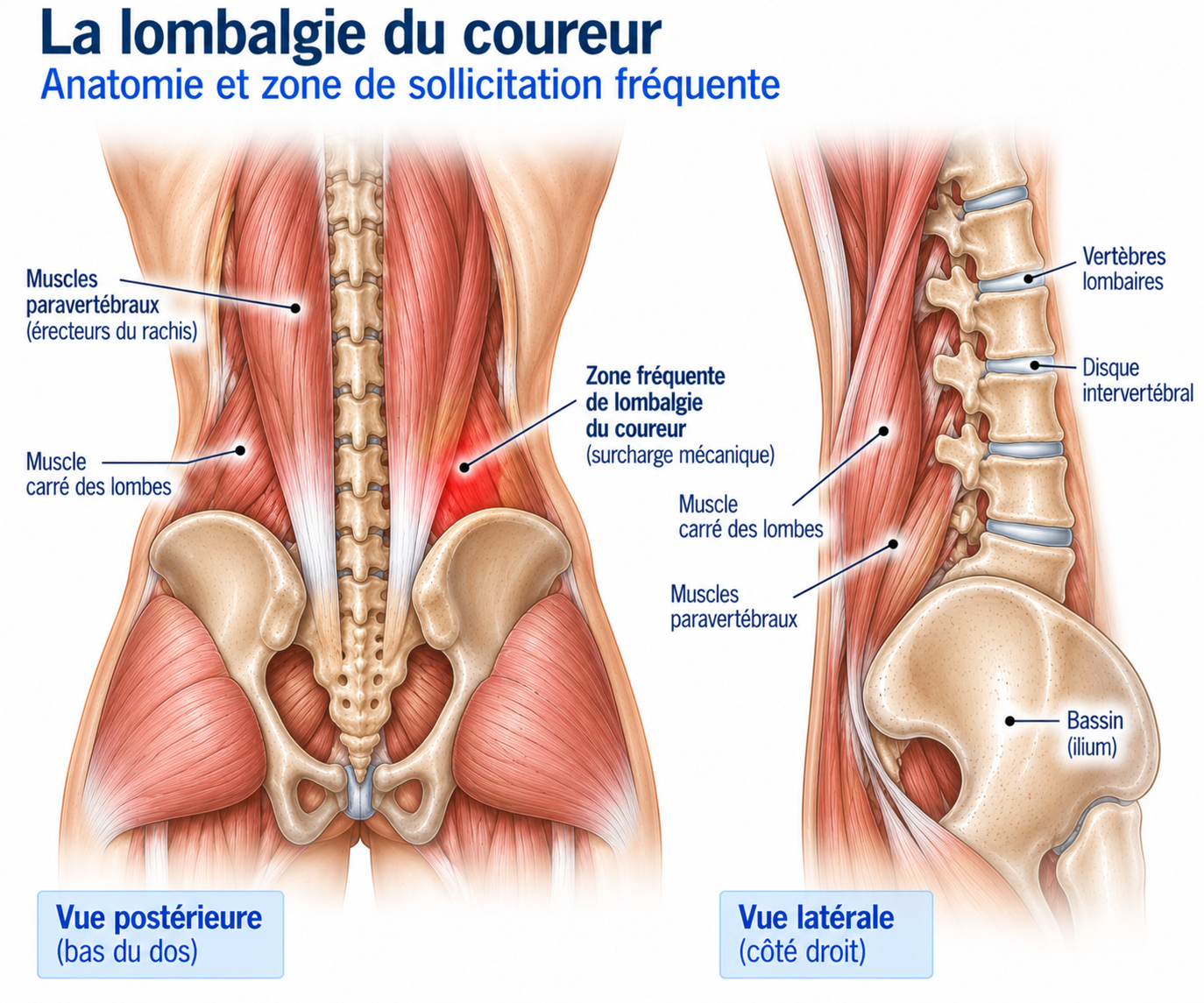
🔍 Click on the image to enlarge it
The intervertebral disc
Between two vertebrae lies an intervertebral disc, made up of a central nucleus pulposus and a peripheral annulus fibrosus, which contributes to shock absorption, mobility and load distribution. With age, it is common to see reduced hydration, tears in the annulus, loss of disc height, or protrusions and herniations — abnormalities that are also common in people without pain. An MRI showing “disc disease” therefore does not necessarily mean that the disc is responsible for the low back pain.
The posterior joints
The zygapophyseal joints, or facet joints, are located at the back of the vertebrae and contribute to stability, movement control and the limitation of certain ranges of motion. They can be involved in some low back pain, particularly when the pain is aggravated by extension and rotation — but a radiological abnormality of the posterior joints is not enough to establish a causal diagnosis.
The trunk muscles
Lumbopelvic stability depends on many muscles: multifidus, erector spinae, transversus abdominis, obliques, rectus abdominis, quadratus lumborum, diaphragm, pelvic floor muscles and gluteal muscles. Their role is not simply to “keep the back straight”: they make it possible to control trunk movements and to transmit forces efficiently between the lower limbs and the pelvis.
Does running damage the back?
No. There is no general reason to consider running a dangerous activity for the lumbar spine. A systematic review published in 2024 found transient disc changes after running, probably linked to compression and a temporary redistribution of water, but longitudinal data did not show any clear harmful effect of running training on disc health. A meta-analysis published in 2026 even suggests that exposure to physical exercise may have favourable or neutral effects on disc health. The question is therefore less “is running damaging my back?” than “what load can my back currently tolerate?”.
Epidemiology
Low back pain is a common reason for runners to seek medical advice. The vast majority of low back pain in athletes is non-specific and progresses favourably with active management. Situations requiring a specific approach (radicular pain, symptomatic disc herniation, spondylolysis, spondylolisthesis, stress fracture, inflammatory disease) are rarer but must be identified, particularly spondylolysis in young athletes with persistent training-related low back pain.
Causes & risk factors
Low back pain can appear when the load imposed temporarily exceeds the adaptive capacity of the lumbopelvic system: a sudden increase in mileage, a return after a long break, marathon preparation, a large increase in elevation gain, more frequent sessions, interval training, general fatigue, insufficient recovery, an abrupt change of terrain, or a major change in shoes or technique. But low back pain can also appear without any obvious increase in load: the pain is therefore not always linked to an identifiable structural injury.
Contributing factors
Training factors: rapid increase in volume, simultaneous increase in volume and intensity, repeated long sessions, lack of recovery, too rapid a return, more frequent hill sessions, abrupt change in practice.
Muscular factors: gluteal strength deficit, quadriceps deficit, weakness of the trunk muscles, poor endurance of the lumbar extensors, single-leg strength deficit.
Mobility factors: limited hip mobility, limited ankle mobility, reduced thoracic mobility. These factors are not, however, necessary causes: a runner can have excellent mobility and still develop low back pain.
Psychosocial factors
Stress, poor sleep, anxiety, fear of movement, catastrophising, worry about a presumed injury or a feeling of loss of control also play an important role in the persistence of some low back pain. This absolutely does not mean that the pain is “psychological” — the pain is real, but it results from the interaction between tissues, the nervous system, context, load, recovery and psychological factors.
Symptoms
The pain of common low back pain can be central, one-sided, two-sided, low lumbar or sometimes slightly gluteal; it can be triggered by running, sitting, bending forward, bending backward, rotation or carrying loads, and can vary from one day to the next. Mechanical low back pain can sometimes be very intense while remaining benign.
Acute low back pain comes on suddenly, sometimes during a movement, a training session, an effort, bending forward or a rotation; the patient may feel “locked”, which often corresponds to an acute pain reaction with reflex limitation of movement rather than a “displaced” vertebra. Most episodes progress favourably. Low back pain is described as chronic beyond three months of symptoms; the pain may then become less directly linked to a specific tissue injury, with possible deconditioning, loss of strength, reduced activity, fear of movement, hypersensitivity, sleep disturbance and a gradual drop in sporting level.
Low back pain associated with pain radiating into the buttock, thigh, leg or foot may correspond to radicular pain, sometimes with pins and needles, numbness, muscle weakness or a reduced reflex. Sciatica, radicular pain in the distribution of the sciatic nerve, is often linked to the L5 or S1 nerve roots — but pain in the buttock or the back of the thigh is not necessarily sciatica and must be distinguished from piriformis syndrome, proximal hamstring tendinopathy, sacroiliac pain or hip pathology.
In young runners, spondylolysis (a stress injury of the pars interarticularis) should be considered in cases of persistent low back pain made worse by extension, localised pain during training and no improvement despite simple management. Spondylolisthesis, the forward slippage of a vertebra relative to the one below, may be isthmic, degenerative, traumatic or dysplastic; its presence on imaging is not necessarily responsible for the pain.
Clinical examination
The examination must be comprehensive: posture, lumbar and hip mobility, strength, sensation, reflexes, neurological testing, gait, pelvic control and single-leg control. A neurological examination is essential when radiating symptoms are present: strength (dorsiflexion, big toe extension, plantar flexion, knee extension and flexion), sensation in the nerve root distributions, patellar and Achilles reflexes, and neural tension tests (Lasègue). A clear or progressive motor deficit is particularly important to identify.
In runners, functional analysis (squat, single-leg squat, step-down, lunge, jump, single-leg balance, running) looks at pelvic control, trunk movement, hip stability, knee control and landing strategy — the runner must be considered as a functional chain.
Warning signs
Prompt medical advice is needed in the event of a significant or progressive motor deficit, saddle anaesthesia, new urinary or sphincter problems, loss of bowel control, weakness in both legs, fever, a marked deterioration in general health, unusual night pain, a history of cancer, immunosuppression, significant trauma or unexplained weight loss. The combination of low back pain with sphincter problems or saddle anaesthesia requires urgent investigation for cauda equina syndrome.
Further tests
X-rays
Useful for looking for spondylolisthesis, a fracture, a bone abnormality, a deformity, significant osteoarthritis or certain abnormalities of the pars interarticularis. Dynamic views can sometimes be used to look for instability. Not routine.
Lumbar MRI
Examines the discs, nerve roots, spinal canal, conus medullaris, bone, posterior joints and soft tissues. Particularly useful in cases of persistent radicular pain, neurological deficit, suspected serious pathology or stress injury, or persistent symptoms with a possible indication for intervention. Acute low back pain without warning signs does not generally require immediate imaging (the American College of Radiology considers X-rays and MRI generally inappropriate in this context). Degenerative abnormalities (protrusions, herniations, disc disease, facet osteoarthritis) are extremely common in people without symptoms: imaging must always be compared with the symptoms, the clinical examination and the course of the condition.
Differential diagnoses
Paravertebral muscles
Localised pain, often linked to load.
Facet joints
Posterior pain, sometimes aggravated by extension and rotation.
Intervertebral disc
Some low back pain is consistent with a discogenic origin.
Nerve root
Radicular pain, possibly with a neurological deficit.
Sacroiliac joint
Buttock or lumbo-gluteal pain.
Hip
An intra-articular problem may be felt in the groin or gluteal region.
Proximal hamstring tendinopathy
Low buttock pain, often in runners.
Spondylolysis
Particularly in adolescents and young athletes.
Stress fracture
To be looked for depending on the context.
Inflammatory disease
To be considered in particular in cases of inflammatory-type low back pain.
Classification
Grade 1 — Mild low back pain
Mild pain, daily activities maintained, running possible, no neurological deficit.
Grade 2 — Symptomatic low back pain
Regular pain, reduced training volume, discomfort after sessions, limitation of some ranges of motion.
Grade 3 — Significant low back pain
Significant limitation of running, persistent pain, reduced daily activities, functional deficit.
Grade 4 — Specialist assessment needed
Neurological deficit, significant persistent radicular pain, suspected spondylolysis or fracture, night pain or red flag, cauda equina syndrome.
Treatment
Controlling the pain flare-up
Modern management of low back pain is based mainly on an active approach: education, staying active, temporary adjustment of load, exercises, strengthening and a gradual return to running. Prolonged strict rest is rarely useful.
During an acute flare-up, activities that strongly trigger symptoms are temporarily reduced (lower mileage, temporary removal of interval training, fewer hills and long runs, lighter heavy loads, adaptation of painful exercises), while maintaining as far as possible walking, daily activity, cycling if pain-free, swimming and suitable exercises. The aim is to move enough without maintaining excessive irritation.
Restoring mobility and strengthening the trunk
Mobility should be worked on without forcing: gentle lumbar mobility (flexion-extension within a comfortable range), controlled rotation (gentle thoracic and lumbopelvic mobilisation), hip mobility according to the deficits identified, and thoracic mobility — particularly useful for some runners with poor thoracic mobility.
Strengthening the trunk is not just about doing sit-ups: front plank (plank, then progression), side plank (obliques and lateral control), bird-dog (alternating arm and leg extension), dead bug (trunk control with limb movements) and Pallof press (anti-rotation work). The aim is to improve the trunk's capacity to resist movement rather than to seek permanent stiffness.
Strengthening the glutes and lower limb
The gluteal muscles play a major role in transmitting forces: glute bridge, hip thrust, abduction, lateral band walk, squat, lunge, deadlift. In runners with chronic low back pain, one randomised trial notably showed that a programme targeting the lower limb could improve specific function more than programmes focused solely on the lumbar extensors or lumbar stabilisation.
The programme should progressively include the quadriceps, hamstrings, glutes, calves and trunk muscles (squat, split squat, lunge, step-up, deadlift, hip thrust, calf raises), progressing by increasing load and volume, reducing rest times and increasing complexity. Single-leg work (balancing on one leg, assisted single-leg squat, step-down, lunge, single-leg hop) aims for pelvic control, trunk stability, knee control and good hip-knee-ankle coordination — since running is essentially a succession of single-leg stances.
Reintroducing impacts and returning to running
Before returning to running: brisk walking, climbing stairs, small jumps, single-leg hops, bounds, plyometric work, then running. Progression depends on tolerance; the runner does not need to be completely symptom-free before each exercise, but a gradual and lasting increase in symptoms should lead to a reduction in load.
The return to running can begin by alternating 1 minute of running / 2 minutes of walking for 20 to 30 minutes, then increasing running time, shortening the breaks, running continuously, increasing distance, and then reintroducing elevation gain and speed. The next-day response can be monitored (24-hour rule): a session is generally acceptable if pain remains under control, does not get progressively worse during the session, is not accompanied by any new neurological symptom and if usual function returns quickly.
Complementary treatments
Medication
Paracetamol, non-steroidal anti-inflammatory drugs or specific treatments for neuropathic pain may be used occasionally alongside active management, depending on age, kidney function and associated risks. They do not replace a gradual return to activity.
Manual therapy
Manipulation, mobilisation or massage can sometimes temporarily reduce pain, as a complement. In athletes, the available data do not establish the superiority of any particular manual technique; they should not make up the whole of the treatment.
Injections
They are not a routine treatment for common low back pain. They may have a place in certain very selected situations (radicular pain, suspected facet pain) but require a precise diagnosis and do not replace rehabilitation.
Surgery
Most common low back pain does not require surgery. It may be discussed in certain situations: significant neurological deficit, cauda equina syndrome, persistent radicular pain with matching compression, certain instabilities or particular structural lesions — imaging must then show an abnormality consistent with the symptoms. A herniated disc visible on MRI in a patient without neurological symptoms is not in itself an indication for surgery.
When a herniated disc causes radicular pain, initial treatment often remains conservative in the absence of a severe neurological deficit, and most patients progress favourably. An MRI becomes particularly relevant when symptoms persist or when a procedure (surgery or epidural injection) is being considered.
Recovery time
There is no universal duration. Most episodes of acute low back pain progress favourably; for common low back pain, the aim is often to maintain or quickly reintroduce suitable activity rather than to wait for a fixed period. Low back pain is described as chronic beyond three months of symptoms. For a specific injury such as spondylolysis, the timeline for returning depends on the diagnosis and how it progresses, and must be adapted to the stage of healing.
Prevention
Preventing recurrence depends less on a perfect “good posture” than on a good capacity to adapt to load: avoid sudden increases in volume and intensity, keep up regular strengthening (quadriceps, glutes, hamstrings, calves, trunk muscles), plan recovery days, make sure you get enough sleep — as lack of sleep can increase pain sensitivity and reduce recovery capacity — and vary your activities (running, cycling, strengthening, walking, swimming).
Frequently asked questions
Can I run with low back pain?
Does running damage the discs?
Does a herniated disc mean I have to stop running?
Do I need an MRI?
Is core training essential?
Can I do trail running?
Can I do strength training with low back pain?
Can low back pain come from the hip?
Is low back pain in a teenage runner a cause for concern?
How long do I need to stop running?
Common mistakes
"I have disc disease, so my disc is damaged": disc abnormalities are common in people without pain.
"You only need to strengthen the lower back": the hip, the pelvis and the whole lower limb contribute to lumbopelvic control.
"You have to stop moving completely": staying suitably active is generally preferable to prolonged rest.
"You need perfect posture to stop having pain": there is no single posture that protects the back from pain.
"You must avoid bending forward": flexion should be gradually reintroduced when needed.
"Abdominal muscles protect the back": the trunk's capacity to control movement and transmit forces is more important than simple abdominal strengthening.
"An MRI is essential": in the absence of a red flag or neurological deficit, it is generally not needed as a first-line investigation.
"Low back pain means you have to stop running": in many situations, running can be continued or gradually resumed.
Key points — Institut Basque du Coureur
Low back pain in runners is not a single diagnosis. Most low back pain is non-specific and progresses favourably with active management. Certain particular situations must nevertheless be looked for: radicular pain, neurological deficit, symptomatic disc herniation, spondylolysis in young athletes, stress fracture, inflammatory disease or, more rarely, infection or tumour. In runners, treatment is based mainly on staying suitably active, gradual load management, strengthening of the trunk, hips and lower limbs, lumbopelvic control work, gradual reintroduction of impacts and a graduated return to mileage and intensity. The aim is not to protect the back indefinitely from all stress, but to gradually restore its capacity to withstand the stresses of running. Finally, an MRI showing disc disease, a protrusion or osteoarthritis does not automatically mean that this abnormality is responsible for the pain: we do not treat an image, we treat a runner, their symptoms, their capacities and their sporting goals.
- Thornton JS, Caneiro JP, O'Sullivan P, et al. Treating low back pain in athletes: a systematic review with meta-analysis. Br J Sports Med. 2021;55:656-663.
- Shu D, Dai S, Wang J, et al. Impact of Running Exercise on Intervertebral Disc: A Systematic Review. Sports Health. 2024;16(6):958-970.
- Samanna CL, et al. The Impact of Exercise on Intervertebral Disc Health: A Systematic Review and Meta-Analysis. Sports Med. 2026;56(4):941-965.
- Wang XQ, Zheng JJ, Yu ZW, et al. A meta-analysis of core stability exercise versus general exercise for chronic low back pain. PLoS One. 2012;7:e42505.
- Searle A, Spink M, Ho A, Chuter V. Exercise interventions for the treatment of chronic low back pain: a systematic review and meta-analysis of randomised controlled trials. Clin Rehabil. 2015;29:1155-1167.
- Van Middelkoop M, Rubinstein SM, Verhagen AP, et al. Exercise therapy for chronic nonspecific low-back pain. Best Pract Res Clin Rheumatol. 2010;24:193-204.
- Aasa U, Berglund L, Michaelson P, Djupsjöbacka M. Individualized low-load motor control exercises and education versus a high-load lifting exercise and education in patients with chronic low back pain. J Orthop Sports Phys Ther. 2015.
- Koldas Dogan S, et al. Comparison of Lower Limb and Back Exercises for Runners with Chronic Low Back Pain. Med Sci Sports Exerc. 2017;49:2108-2116.
- Hayden JA, Ellis J, Ogilvie R, Malmivaara A, van Tulder MW. Exercise therapy for chronic low back pain. Cochrane Database Syst Rev. 2021;9:CD009790.
- Chou R, Qaseem A, Owens DK, Shekelle P. Diagnostic imaging for low back pain: advice for high-value health care. Ann Intern Med. 2011;154:181-189.
- American College of Radiology. ACR Appropriateness Criteria: Low Back Pain. Updated guidance.
- Koes BW, van Tulder MW, Thomas S. Diagnosis and treatment of low back pain. BMJ. 2006;332:1430-1434.
- Foster NE, Anema JR, Cherkin D, et al. Prevention and treatment of low back pain: evidence, challenges, and promising directions. Lancet. 2018;391:2368-2383.
- Mazzola A, et al. Spondylolysis and spondylolisthesis in athletes: diagnosis and management. J Sports Med Phys Fitness.
- Selhorst M, Fischer A, Ravindran R, et al. Rehabilitation considerations for spondylolysis in adolescent athletes. J Orthop Sports Phys Ther.