
Definition
Popliteal artery entrapment syndrome (SAPP in French), or Popliteal Artery Entrapment Syndrome (PAES), is a rare vascular condition in which the popliteal artery is abnormally compressed by the surrounding muscular or tendinous structures, most often at the level of the medial head of the gastrocnemius muscle. This compression may be dynamic (appearing mainly during calf contraction), permanent (related to an anatomical anomaly), or progressively responsible for arterial lesions: stenosis, thrombosis, aneurysm or occlusion.
The syndrome mainly affects young, athletic individuals without classic cardiovascular risk factors. In runners, it typically presents as calf pain that is reproducible on exertion, sometimes accompanied by a feeling of tightness, cramping, paraesthesia or reduced performance.
The main problem is that this presentation closely resembles that of other, much more common causes of exertional leg pain, in particular chronic exertional compartment syndrome. The diagnosis should therefore be considered when symptoms are highly reproducible, atypical, or resistant to standard muscle-focused management.
Anatomy
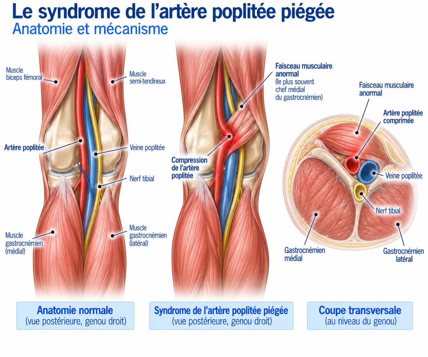
The popliteal artery is the continuation of the femoral artery after it passes behind the knee. It runs through the popliteal fossa, a space located between the femur, the joint capsule, the popliteus muscle, the medial and lateral gastrocnemius muscles, and the popliteal nerve and venous structures (tibial nerve, popliteal vein).
In a normal anatomical situation, the popliteal artery passes close to the medial head of the gastrocnemius, but without being significantly compressed when the muscle contracts. In some patients, the anatomical relationship between the artery and the gastrocnemius is abnormal: the artery may then be displaced, medialised, compressed by an abnormal muscle attachment, surrounded by a fibrous band, or compressed against bony structures during ankle movements.
🔍 Click on the image to enlarge it
It is this anatomical variation — or, in functional forms, gastrocnemius hypertrophy during exercise — that explains why calf contraction can, in some athletes, compress the popliteal artery transiently or permanently.
Epidemiology
Popliteal artery entrapment syndrome is rare in the general population, but its frequency is probably underestimated in young athletes with exertional claudication. It mainly affects young adults and sportspeople and endurance athletes — runners, footballers, military personnel, people practising sports that place heavy demands on the calf — usually without atherosclerosis or classic cardiovascular risk factors.
The presentation may be bilateral, which is particularly common in functional forms: both lower limbs should therefore be examined systematically when PAES is suspected.
There is an important difference between true anatomical compression and functional compression related solely to muscle contraction, with no obvious congenital anatomical anomaly — this distinction directly influences the treatment strategy.
Causes & risk factors
Compression mechanism: the Whelan classification
The classic mechanism involves an abnormality in the development or position of the medial gastrocnemius. The historical Whelan classification distinguishes several anatomical forms:
Type I: the popliteal artery follows an abnormal course in relation to the medial head of the gastrocnemius.
Type II: the medial head of the gastrocnemius has an abnormal attachment, causing deviation or compression of the artery.
Type III: a muscular or accessory band compresses the artery.
Type IV: the artery is compressed by the popliteus muscle or another deep structure.
Type V: a combination of several anomalies, with possible involvement of the popliteal artery and/or vein.
Type VI (functional form): there is not necessarily any obvious congenital anatomical anomaly. Compression occurs mainly during muscle contraction — plantar flexion, gastrocnemius contraction, intense effort — particularly in athletes with especially well-developed calves. This form is particularly important in runners.
Anatomical or functional form?
This distinction is essential. In the anatomical form, the artery is compressed by a structural anomaly and may progressively develop stenosis, intimal injury, thrombosis, post-stenotic aneurysm or even occlusion; management is generally surgical when the compression is symptomatic. In the functional form, the artery is anatomically relatively normal at rest and compression appears mainly during exercise; this form, which is harder to diagnose, must in particular be distinguished from chronic exertional compartment syndrome, muscle impingement, a calf strength/endurance deficit or a biomechanical problem of the lower limb. Recent data suggest that some selected functional forms can be managed conservatively, whereas symptomatic anatomical forms are more likely to require surgical decompression — the level of evidence nevertheless remains limited.
Pathophysiology
During running, gastrocnemius contraction increases. In a patient with PAES, this contraction can cause transient compression of the artery, with reduced distal blood flow. Early in the disease, this obstruction is purely functional: the patient may have a normal vascular examination at rest, normal pulses, a normal ankle-brachial index and normal standard imaging. During exercise, however, the reduced flow can cause exertional muscle ischaemia, which is responsible for the pain. In the long term, repeated compression can also damage the arterial wall.
Why does running trigger the symptoms?
Running combines several factors that promote compression: repeated gastrocnemius contraction, high muscular metabolic demand, increased blood flow required by the calf, repeated plantar flexion movements, high speed, hills, accelerations and running on the toes. A patient may therefore be symptom-free when walking, at rest or during normal daily activity, yet develop symptoms after a few minutes of running. A reproducible distance or time before the onset of symptoms is a particularly useful element in guiding the diagnosis.
Contributing factors
Several situations should raise diagnostic suspicion.
Profile: young athlete, intensive training, no cardiovascular risk factors, bilateral symptoms.
Sport: running, sprinting, football, sports that place heavy demands on the calf or involve many accelerations.
Muscle morphology: marked gastrocnemius hypertrophy may contribute to the phenomenon in some functional forms.
History: persistent calf pain despite properly conducted management of compartment syndrome or a muscle injury should also prompt reconsideration of the diagnosis.
Symptoms
The classic picture is intermittent claudication in an athlete. The patient describes exertional calf pain, tightness or a feeling of compression, cramping, heaviness, unusual calf fatigue, reduced power, and sometimes paraesthesia or numbness in the foot. The pain usually appears after a relatively reproducible time or distance, and disappears once exercise stops.
The pain is usually located in the calf, sometimes deep in the back of the lower leg, sometimes at the junction of the upper and middle thirds of the calf. It can be difficult to distinguish from muscle pain: it is the combination with the other features that should point towards a vascular origin.
Certain features are particularly suggestive in runners: pain that almost always appears after a similar duration of exercise, rapid resolution on stopping, inability to reproduce the pain by palpating the muscle, no muscle abnormality on imaging, bilateral symptoms, a feeling that the calf “locks up” during exercise, associated paraesthesia, reduced performance with no muscular explanation, and symptoms triggered mainly by hills or accelerations.
A patient may, for example, report: “I can run for 15 minutes, then my calf becomes very painful. If I stop for a few minutes, I can set off again.” This notion of a reproducible onset threshold is particularly useful in guiding the diagnosis.
Clinical examination
The clinical examination should be performed at rest and then after a provocative manoeuvre. The examination at rest may be completely normal: the dorsalis pedis and posterior tibial pulses, skin temperature, colour, capillary refill time, pulse symmetry, trophic status and any vascular scars should be assessed. Palpation of the muscles also looks for pain reproduced at the gastrocnemius.
Dynamic examination is an essential part of the diagnosis: the aim is to detect a change in pulses or arterial flow during resisted plantar flexion, forced dorsiflexion, gastrocnemius contraction, in the standing position or during exercise that reproduces the symptoms. Loss or reduction of the arterial signal during a provocative manoeuvre should raise suspicion of compression — but be careful: reduced flow during a provocative manoeuvre is not enough on its own to make the diagnosis, as some compressions can be observed in asymptomatic individuals. Interpretation must therefore always be correlated with the symptoms, the examination, imaging and the sporting context.
Normal imaging at rest does not rule out the diagnosis
In a functional form, the artery can be completely normal at rest, with no stenosis and normal flow: compression appears only when the gastrocnemius contracts. A purely static assessment can therefore miss the diagnosis; in an athlete with strong clinical suspicion, a dynamic assessment reproducing the symptoms or the suspected mechanism should be preferred.
Further tests
Ankle-brachial systolic pressure index (ABI)
May be normal at rest; a normal ABI therefore does not rule out dynamic PAES. It is more useful when measured after exercise, immediately after a provocative manoeuvre, or in comparison with the contralateral limb. A significant drop in distal pressure after exercise strengthens the suspicion of a functional arterial limitation.
Doppler ultrasound
Generally one of the first investigations performed. It assesses the popliteal artery, blood flow velocities, the flow profile, the possible presence of stenosis and changes in flow during manoeuvres. The examination should ideally be dynamic: at rest, in plantar flexion, in dorsiflexion, during calf contraction and possibly after exercise. It can also assess the muscle structures responsible for the compression.
Dynamic CT angiography
Performed with acquisitions comparing a resting position and a provocative position (sometimes with forced plantar flexion). It allows analysis of the gastrocnemius anatomy, the course of the artery, the level of compression, the degree of stenosis, any occlusion and associated arterial lesions. Particularly informative in functional forms.
MR angiography
A useful alternative when it is preferable to limit radiation exposure. It assesses vascular anatomy, muscles, soft tissues and any associated lesions; particularly useful when a muscular or neurological differential diagnosis remains possible.
Arteriography
Much less used today as an initial diagnostic investigation. It may be performed in certain complex situations: significant arterial lesion, thrombosis, surgical planning or persistent diagnostic doubt.
Differential diagnoses
Chronic exertional compartment syndrome
The main differential diagnosis: it also causes reproducible pain, calf tightness, symptoms appearing after a certain duration of exercise and disappearing once exercise stops, sometimes with paraesthesia. The distinction is based on vascular investigations and, when indicated, measurement of intramuscular pressures.
Muscle injury (gastrocnemius or soleus)
Usually causes localised pain that is tender on palpation, painful on contraction and sometimes on stretching. Ultrasound or MRI can often identify it.
Tibial stress fracture
To be considered in the case of localised pain that persists after exercise, is painful on bone palpation and progresses over time.
Shin splints / medial tibial stress syndrome
The pain is usually more diffuse and medial along the tibia.
Nerve involvement
Nerve compression can cause paraesthesia, numbness, weakness or radiating pain.
Atherosclerotic arterial claudication
Much rarer in young athletes, but should be considered depending on the patient's profile.
Venous disease
Venous thrombosis or chronic venous disease can cause pain or a feeling of tightness, but the clinical picture is different.
Classification
Level 1 — Low suspicion
Pain that is not very reproducible, with no obvious vascular features. Look first for a muscular, tendinous or bone cause.
Level 2 — Intermediate suspicion
Pain that is highly reproducible on exertion, with no obvious muscle abnormality. Carry out a dynamic vascular assessment.
Level 3 — High suspicion
Reproducible pain associated with neurological or vascular symptoms and a change in flow during manoeuvres. Specialist vascular assessment with dynamic imaging.
Level 4 — Confirmed PAES
Documented arterial compression, correlated with symptoms, with or without arterial lesion. Vascular surgery opinion.
Treatment
Management according to the form (anatomical or functional)
Management depends mainly on the form identified. In a symptomatic anatomical form, it is generally directed towards specialist vascular care, most often surgical. In a functional form, the decision is individualised between load modification, rehabilitation, investigation for an associated condition (in particular compartment syndrome) and surgical treatment reserved for persistent symptomatic forms. Recent literature stresses that the data remain heterogeneous and of low certainty, particularly for functional forms.
Phase 1 – Reduce vascular stress
When the diagnosis is suspected but the situation does not require urgent intervention, the aim is to temporarily reduce the triggering efforts: avoid sprints, limit hills, reduce running on the toes and favour activities that do not reproduce the symptoms. The aim is not necessarily to stop sport completely, but to avoid situations that systematically cause exertional ischaemia.
Phase 2 – Rehabilitation
In selected functional forms, rehabilitation may be considered. It includes mobility work (ankle mobility, progressive dorsiflexion, mobility of the ankle-foot complex), muscle work (soleus, gastrocnemius, tibialis anterior, fibular muscles, intrinsic foot muscles) and proximal work (gluteus medius, gluteus maximus, pelvic control, single-leg stability), in order to reduce excessive stress on the calf and improve overall biomechanical efficiency.
Calf strengthening should be progressive: double-leg calf raises, then single-leg, soleus work with the knee bent, loaded eccentric and concentric work, plyometrics, then running with accelerations and changes of pace. In a patient with documented functional vascular compression, this strengthening should be carried out with caution and ideally as part of specialist care.
There is no evidence to support gastrocnemius stretching as a specific treatment for PAES: it may improve mobility when there is an associated limitation, but calf stiffness should not be regarded as the main cause of an objectively confirmed vascular problem — an aggressive stretching programme does not correct an anatomical anomaly in the course of the artery.
Return to running
Return to running depends on the mechanism identified. In a functional form managed conservatively, you can start with symptom-free brisk walking, then alternate 2 minutes of running and 2 minutes of walking, before gradually increasing running time up to continuous running. Distance is then increased, followed by the gradual introduction of hills and finally accelerations. Speed and intensity should be reintroduced last.
Criteria for returning to sport
Return should ideally combine no pain during usual exercise, no paraesthesia, no abnormal feeling of tightness, good calf strength and endurance, no functional limitation and, when follow-up investigations are indicated, no worrying vascular abnormality. In elite athletes, return should be individualised with the vascular surgeon and the sports physician.
Surgery
When to refer to a vascular surgeon?
A specialist consultation is particularly indicated in the case of calf claudication in a young athlete, symptoms reproducible on exertion, clinical suspicion of arterial compression, an abnormality on dynamic Doppler, a drop in ABI after exercise, popliteal stenosis or occlusion in a young person, unexplained popliteal thrombosis, a popliteal aneurysm in a young person, or failure of management for presumed compartment syndrome.
When is surgery needed?
Surgery is generally considered when symptomatic anatomical compression has been demonstrated, when symptoms significantly limit activity, when there is an arterial lesion, significant stenosis, thrombosis or occlusion, or when conservative treatment is insufficient in a selected functional form. The aim is to release the artery and remove the structure responsible for the compression.
Surgical decompression
Surgery may involve release of the popliteal artery, division of an abnormal muscular or tendinous structure, myotomy, partial resection of the gastrocnemius in certain functional forms, and an associated fasciotomy when compartment syndrome is also present. In functional forms in athletes, some teams use targeted resection of part of the medial head of the gastrocnemius to eliminate dynamic compression; the results published in specialist case series are encouraging, but the studies remain mainly retrospective.
PAES and chronic exertional compartment syndrome can coexist: a patient may have popliteal compression, compartment syndrome, or both. In series of athletes operated on for a functional form, a fasciotomy combined with popliteal decompression is frequently performed when compartment syndrome is suspected or documented — it is therefore important not to systematically set the two diagnoses against each other.
Does the artery need to be repaired?
Not always. If the artery is simply compressed but remains healthy, decompression may be sufficient. If repeated compression has led to significant stenosis, intimal injury, thrombosis, aneurysm or occlusion, arterial reconstruction may become necessary, in particular using an autologous vein segment. Early diagnosis is therefore important: treating compression before an arterial lesion develops potentially allows simpler surgery.
Rehabilitation after surgery
Recovery depends on the procedure performed and above all on the condition of the artery. It generally follows several phases: a protection phase (wound healing, recovery of mobility, pain control, resumption of walking), a muscle recovery phase (ankle mobility, calf activation, quadriceps, hamstrings, gluteal muscles), a strengthening phase (single-leg work, calf raises, squats, lunges, soleus work, proprioceptive exercises), an impact phase (double-leg then single-leg jumps, bounds, plyometric work, changes of direction), and finally a return to running phase (brisk walking, walk-run intervals, continuous running, increased volume, hills, accelerations, interval training, return to sport-specific activity).
Recovery time
Results are generally favourable but vary according to the form and the initial condition of the artery. A recent series of athletes operated on for a functional form reported symptom improvement in around 86% of limbs and a return to the previous level of sport in around 82% of athletes, with delayed recurrence of symptoms observed in a minority of cases. Another specialist series reported a return to the previous competitive level in around 78% of athletes.
A technically released artery does not automatically mean an immediate return to the previous level of sport: muscular, vascular and functional recovery may take several weeks to several months.
The vascular prognosis after surgery is generally good: long-term series report satisfactory patency rates after decompression and, when necessary, arterial reconstruction — one five-year follow-up study found a patency rate of 84% after surgery, in a population that included forms with arterial involvement requiring reconstruction. Functional results, however, remain more variable: a distinction must be made between anatomical success, resolution of symptoms, functional recovery and return to the previous level of sport — these four elements are not necessarily equivalent.
Several mechanisms can explain persistent symptoms after surgery: long-standing arterial damage, associated compartment syndrome, muscle deconditioning, calf strength deficit, pain sensitisation, poor recovery after a long period of reduced activity, or another associated diagnosis. Recurrence of symptoms should also prompt a search for new compression, residual compression, compartment syndrome, a vascular problem or another cause of exertional pain.
Prevention
There is no strategy to prevent a congenital anatomical anomaly. However, in athletes with a functional form, certain measures can limit stress: a well-planned training progression, avoiding sudden increases in volume; maintaining good ankle mobility; progressive strengthening of the calf and posterior chain; varying surfaces and types of loading; and avoiding systematically running at high intensity when symptoms are present.
Frequently asked questions
Can you have popliteal artery entrapment syndrome with normal pulses?
Is it a dangerous condition?
Can I keep running?
Is it the same thing as compartment syndrome?
Is a Doppler scan enough to make the diagnosis?
Does a normal MRI rule out the diagnosis?
Is surgery always necessary?
Can you return to sport after surgery?
Can the syndrome be bilateral?
Should calf pain that occurs only on hills prompt investigation for PAES?
Common mistakes
Immediately assuming a muscle problem: calf pain in a runner is not necessarily muscular.
Diagnosing compartment syndrome without confirmation, even though the clinical picture can be extremely similar. When treatment does not work, the diagnosis must be reassessed.
Being reassured by normal pulses at rest, even though in a functional form pulses can be completely normal at rest.
Performing only static imaging, even though dynamic compression can disappear completely at rest.
Regarding any provoked compression as pathological, even though reduced flow during a manoeuvre can be observed in asymptomatic individuals: the result must be correlated with the symptoms and the full work-up.
Forgetting the possibility of bilateral involvement: assessing the contralateral side may be relevant, particularly in functional forms.
Forgetting associated compartment syndrome: the two conditions can coexist.
Key points — Institut Basque du Coureur
Popliteal artery entrapment syndrome is a rare but important cause of exertional calf pain in young athletes. It should be considered in particular in the case of highly reproducible pain that appears after a relatively constant duration of exercise, disappears quickly at rest, is associated with bilateral symptoms or paraesthesia, has no muscular explanation, or when management for compartment syndrome has failed. The diagnosis relies on a dynamic approach, because the artery can be completely normal at rest: Doppler ultrasound with provocative manoeuvres is often a first-line investigation, supplemented if necessary by dynamic vascular imaging. Anatomical forms must be distinguished from functional forms: in symptomatic anatomical forms, surgical decompression is generally the reference treatment, whereas in functional forms the decision must be individualised, in particular by looking for associated compartment syndrome. Finally, in runners, treatment success should not be judged solely on vascular imaging: resolution of claudication, muscle recovery and return to the previous level of sport are the true goals.
- Cui HJ, Wu YF. Popliteal Artery Entrapment Syndrome: A Systematic Review of Contemporary Management and Outcome Reporting. J Endovasc Ther. 2026.
- Cabot JH, Yang L, Shalita C, et al. Midterm outcomes from a 20-year experience treating functional popliteal artery entrapment syndrome in athletes. J Vasc Surg. 2025;82(3):932-940.
- Levien LJ. Popliteal artery entrapment syndrome. Semin Vasc Surg. 2003;16(3):223-231.
- Gourgiotis S, Aggelopoulos D, Salemis N, Elias C, Brunaud L. Diagnosis and surgical approach of popliteal artery entrapment syndrome: a review. J Vasc Surg. 2006;44(5):1055-1062.
- Lejay A, Delay C, Georg Y, et al. Five Year Outcomes of Surgical Treatment for Popliteal Artery Entrapment Syndrome. Eur J Vasc Endovasc Surg. 2016;51(4):557-564.
- Turnipseed WD. Functional popliteal artery entrapment syndrome: a potentially debilitating vascular condition in young athletes. J Vasc Surg. 2009;49(5):1189-1195.
- Hislop M, Kennedy D, Cramp B, Dhupelia S. Functional popliteal artery entrapment syndrome: a review of the literature and a case report. J Sports Med Phys Fitness. 2014;54(6):738-744.
- Torabi M, Ponsford S, et al. Functional popliteal artery entrapment syndrome in athletes: diagnosis and management. J Vasc Surg. 2019.
- Functional Outcome After Surgery for Popliteal Artery Entrapment Syndrome. EJVES Vasc Forum. 2026;65:75-82.
- Society for Vascular Surgery. Midterm outcomes from a 20-year experience treating functional popliteal artery entrapment syndrome in athletes. 2025.
- Whelan TJ, et al. Popliteal artery entrapment syndrome: anatomical classification and surgical management.
- Popliteal Artery Entrapment Syndrome: Long-term results of surgical treatment. J Med Vasc. 2024.
Similar injuries
Shin splints (medial tibial stress syndrome)
Inflammation of the membrane covering the tibia caused by repeated impacts, affecting 13 to 17% of runners.
Tibial stress fracture
A tibial micro-fracture caused by repeated stress, requiring a mandatory break from running to allow the bone to heal.
Compartment syndrome
Increased muscle pressure during exercise that can compromise blood flow and nerve function.