
Definition
Morton’s neuroma is a painful condition of the plantar interdigital nerve, most often located between the heads of the 3rd and 4th metatarsals. The 2nd space may also be involved, whereas involvement of the 1st or 4th space is unusual and should prompt a search for another diagnosis.
Despite its name, it is not a true tumour of the nerve. The term actually refers to a compressive and irritative neuropathy: the common plantar digital nerve gradually thickens and becomes fibrotic as a result of repeated mechanical compression between the metatarsal heads. This produces perineural fibrosis, nerve thickening, degenerative changes and often an associated intermetatarsal bursitis — hence the terms Morton’s metatarsalgia or interdigital neuropathy that are sometimes used.
The typical clinical result is a burning or electric pain in the forefoot, sometimes accompanied by tingling or numbness in the toes, triggered by weight-bearing, narrow footwear or running.
Anatomy
In each intermetatarsal space, the common plantar digital nerve runs between the metatarsal heads before dividing into two proper digital nerves supplying sensation to two adjacent toes. The 3rd intermetatarsal space, between the 3rd and 4th metatarsals, is particularly exposed to mechanical stress: the nerve runs there through a narrow anatomical environment, in contact with the deep transverse metatarsal ligament, which links the metatarsal heads together, as well as with the plantar and interosseous structures.
A small intermetatarsal bursa is also present in this space. Bursitis of this bursa can cause pain very similar to that of the neuroma, be associated with it, or even be its true origin — which explains the value of ultrasound in distinguishing between the two mechanisms.
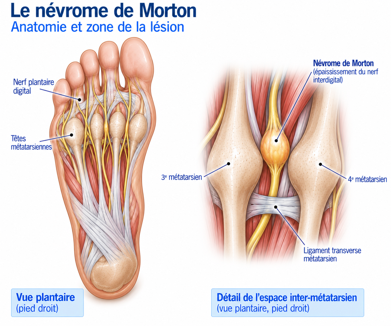
🔍 Click on the image to enlarge it
Why is the 3rd space most often affected?
Several anatomical features explain this predominance: the high mechanical stress on the forefoot at this point, the proximity of the metatarsal heads, their relative mobility, the particular course of the interdigital nerve at this level and the stresses exerted by the surrounding ligamentous structures. The 2nd space may also be involved, but involvement of the 1st or 4th space is unusual and should suggest another diagnosis.
Forefoot biomechanics in running
The problem can be summed up simply: the nerve tolerates repeated mechanical stress poorly when it exceeds its capacity to adapt. During running, the forefoot is subjected to compressive and shear forces, increased pressure under the metatarsal heads, dorsiflexion of the metatarsophalangeal joints and tensioning of the plantar structures. These stresses increase with narrow or heeled footwear, a recent increase in mileage or in the proportion of fast sessions, hills, trail running or jumps. The neuroma can therefore appear after a change in volume, speed, terrain or footwear.
Epidemiology
Morton’s neuroma is a common cause of forefoot pain (metatarsalgia). It mainly affects adults, more often women, as well as people exposed to tight footwear or activities that place heavy demands on the forefoot — runners, people who walk a lot, and sports involving repeated push-offs.
It can be unilateral or bilateral, and several spaces can be affected in the same person. An important point to know: the presence of a neuroma on imaging does not necessarily mean that it is responsible for the symptoms — the diagnosis must always be correlated with the clinical findings.
Causes & risk factors
Footwear
This is one of the most important factors: shoes that are narrow or pointed at the front, high-heeled shoes, stiff shoes that limit the natural mobility of the forefoot, and direct compression of the metatarsals.
Running
A sudden increase in mileage, training frequency, interval sessions, sprints, hills or trail running, a change of shoes or too rapid a transition to minimalist shoes can trigger or perpetuate the symptoms.
Morphology and biomechanics
A cavus foot (a major factor, owing to a very high arch), a flat foot with overpronation, a wide forefoot, a forefoot deformity (hallux valgus), limited ankle dorsiflexion (gastrocnemius tightness), weakness of the intrinsic foot muscles or impaired push-off all promote mechanical overload of the forefoot.
Symptoms
The characteristic symptom is forefoot pain, described as burning, an electric shock, a current-like sensation or tingling radiating to the toes. Some patients report a very suggestive sensation: “it feels like I have a stone in my shoe” or “as if something were stuck under my foot”.
In runners, symptoms tend to appear after several kilometres, when accelerating, running uphill or changing direction, sometimes forcing them to stop, loosen the shoe or massage the forefoot — a very characteristic sign of the condition.
Clinical examination
The diagnosis is primarily clinical. Palpation of the affected intermetatarsal space usually reproduces the usual pain. Transverse compression of the forefoot can provoke the pain and its radiation to the toes, sometimes accompanied by a Mulder’s sign: a palpable or audible click during compression, which is highly suggestive of the diagnosis — although its absence does not rule it out. The thumb-index finger compression test of the intermetatarsal space is also useful: when negative, it makes it reasonable to rule out the diagnosis in an appropriate clinical context.
Further tests
X-rays
These are mainly used to rule out other causes of metatarsalgia (osteoarthritis, fracture, hallux valgus, Freiberg's disease). The neuroma itself is generally not visible on standard X-rays.
Ultrasound
Looks for thickening of the nerve, an associated intermetatarsal bursitis or several neuromas, and allows a dynamic examination of the intermetatarsal space. It can also be used to guide an injection precisely.
MRI
Useful when the diagnosis is uncertain, when several spaces are involved, before surgery is considered in a complex situation, or to look for a differential diagnosis (plantar plate injury, stress fracture).
Differential diagnoses
Mechanical metatarsalgia
Pain under the metatarsal heads, generally less neuropathic, without marked electric shocks or tingling.
Intermetatarsal bursitis
Clinical picture very similar to that of the neuroma, sometimes associated with it — ultrasound often makes it possible to tell them apart.
Plantar plate injury
Particularly common at the 2nd ray; mechanical plantar pain without a typical neurological component.
Metatarsal stress fracture
Gradual pain linked to a recent increase in load, localised more on the bone than between the metatarsals.
Freiberg's disease
Osteonecrosis of the metatarsal head, typically of the 2nd metatarsal, visible on imaging.
Other diagnoses to consider
Metatarsophalangeal arthropathy or capsulitis, sesamoid disorders (pain under the 1st metatarsal), hallux valgus, or more proximal nerve irritation (tibial nerve, peripheral neuropathy).
Classification
Stage 1
Pain only after activity, disappearing quickly with rest. Few or no neurological symptoms, with no significant limitation of running.
Stage 2
Burning or shooting pain during running, sometimes requiring the shoe to be loosened. Discomfort during long walks and reduced tolerance of training volume.
Stage 3
Pain appearing quickly during exercise, symptoms present in daily life, frequent paraesthesia and significant limitation of footwear choice and running.
Stage 4
Symptoms persisting despite well-conducted adjustment of footwear and load, rehabilitation and medical treatment — interventional or surgical treatment may then be discussed.
Treatment
Changing footwear (the key treatment)
The first step, often the most effective, is to reduce mechanical compression of the forefoot. This means choosing a shoe with a wide forefoot, giving the toes enough room, with no lateral compression and a moderate heel. Narrow, pointed or high-heeled shoes should be avoided for the time being.
A foot orthosis with a metatarsal pad may be offered when forefoot mechanics contribute to the symptoms. The pad should be placed proximal to the metatarsal heads, not directly under the painful area: the aim is to distribute pressure better rather than to press on the neuroma — a common mistake that can actually worsen the compression.
Adjusting sporting load and controlling symptoms
It is not necessary to stop all activity systematically: the aim is to determine the level of load that is tolerated. During the painful phase, it is helpful to temporarily reduce mileage, speed, interval training, hills, technical trail running, jumps and long sessions that place heavy demands on the forefoot. Cycling or other non-impact activities can often be continued if they do not reproduce the symptoms.
Restoring foot mobility and function
Once the pain is better controlled, rehabilitation aims to restore mobility of the ankle (dorsiflexion, first ray mobility) and toes, and to strengthen the intrinsic foot muscles — particularly through short foot exercises and active control of the arch. Strengthening of the triceps surae (bilateral then single-leg heel raises) completes this work.
Strengthening, push-off and gradual return to running
Once mobility and strength are satisfactory, more specific work is introduced: single- and double-leg heel raises, single-leg balance exercises, step-ups, then gradually small hops, single-leg hops, skipping rope and changes of direction.
The return to running should be gradual, following the rule duration → distance → frequency → speed: for example, alternating running and walking in short sessions before gradually increasing continuous running time. Progression should be guided by the foot's response in the 24 hours following exercise, rather than by a rigid progression rule.
Before a full return, several criteria must be met: pain-free walking, forefoot compression well tolerated, single-leg exercises and heel raises performed without pain, small hops tolerated and no worsening of symptoms the next day.
Complementary treatments
Corticosteroid injection
May be offered when mechanical measures are not enough, ideally under ultrasound guidance to better target the structure responsible. It does not "repair" the nerve but aims to reduce irritation and pain; repeated injections should be avoided (risk of fat pad atrophy and skin depigmentation).
Shockwave therapy
May be discussed as an adjunct in some persistent forms, but are not a first-line treatment for this indication.
Radiofrequency / cryoablation
Percutaneous techniques discussed in some persistent, resistant forms. There are encouraging data, particularly for radiofrequency, but the studies remain mainly observational and heterogeneous.
Alcohol injections
A specialist option with a variable level of evidence — to be reserved for selected situations, not as a first-line treatment.
Surgery
Surgery is reserved for persistently disabling forms with a reliable clinical diagnosis that are resistant to well-conducted conservative treatment (appropriate footwear, load adjustment, rehabilitation, injections). It should not be decided solely on the basis of the size of the neuroma on imaging: a small neuroma can be very symptomatic, and vice versa.
There are two main strategies. Neurolysis (decompression) aims to free the nerve without removing it, sometimes combined with a transposition; it carries a lower risk of postoperative neurological symptoms. Neurectomy involves removing the diseased portion of the nerve: it results in permanent loss of sensation in the corresponding digital territory, but often brings a clear improvement in pain. However, the quality of studies comparing these two approaches remains limited.
The operation can be performed via a dorsal approach (the most common, making access to the intermetatarsal space easier) or via a plantar approach, used in some revision surgeries but carrying a greater risk of a painful scar under the weight-bearing area.
Recovery time
After straightforward surgery, walking is generally resumed gradually according to the surgeon’s instructions, in three main phases: pain control and wound healing, then prolonged walking, strengthening and balance work, then a gradual return to impact activities and running.
The return to sport should be guided by symptoms and wound healing rather than by a fixed date, progressing from walking to continuous running and then to accelerations and interval training. Some residual tenderness in the forefoot may persist for several weeks or even several months, without compromising the overall result.
Prevention
Prevention relies mainly on reducing the mechanical stress placed on the forefoot. The choice of footwear is essential: favour a wide toe box, no lateral compression and a sole suited to the shape of the foot.
In terms of training, sudden increases in mileage, speed, elevation gain or sessions that place heavy demands on the forefoot should be avoided. Regular strengthening of the intrinsic foot muscles and the triceps surae, together with maintaining single-leg control, ankle mobility and push-off capacity, complete this prevention.
Frequently asked questions
Is Morton's neuroma serious?
Can I run with Morton's neuroma?
Do I need to stop sport?
Can it heal without surgery?
Can the neuroma go away?
Can the neuroma be bilateral?
Why does it only hurt with certain shoes?
Is an ultrasound scan mandatory?
Should a neuroma seen on MRI be operated on?
After a neurectomy, do you lose sensation in the toes?
Can I return to running after surgery?
Common mistakes
Massaging the neuroma firmly, when repeated compression can in fact sustain the irritation.
Stopping running completely, when a temporary reduction in load is often enough.
Placing the metatarsal pad directly under the pain, which can increase pressure instead of relieving it.
Thinking that a neuroma must be large on ultrasound to be painful — size does not predict the intensity of symptoms.
Believing that a normal MRI rules out the diagnosis, when it remains first and foremost clinical.
Thinking that an effective injection solves the problem for good if the footwear issue or mechanical overload persists.
Key points — Institut Basque du Coureur
Morton's neuroma is most often a compressive neuropathy of the interdigital nerve of the forefoot, usually in the 3rd intermetatarsal space. It causes burning or electric pain, sometimes accompanied by tingling and a feeling of a foreign body in the shoe. The diagnosis is primarily clinical; ultrasound is particularly useful to confirm it, look for an associated bursitis and guide a possible injection. Treatment always begins by reducing mechanical stress — appropriate footwear and management of sporting load — before considering, in the event of prolonged failure, an injection and then, more rarely, surgery.
- Mak MS, Chowdhury R, Johnson R. Morton's neuroma: review of anatomy, pathomechanism, and imaging. Clin Radiol. 2021;76(3):235.e15-235.e23.
- Pitcher M, Moulson A, Pitcher D, Herbland A, Cairns MC. Diagnostic Accuracy of Subjective Features and Physical Examination Tests for Morton Neuroma: A Systematic Review. Foot Ankle Orthop. 2024;9(4):24730114241291055.
- Matthews BG, Thomson CE, Harding MP, McKinley JC, Ware RS. Treatments for Morton's neuroma. Cochrane Database Syst Rev. 2024;2(2):CD014687.
- Xu Z, Duan X, Yu X, Wang H, Dong X, Xiang Z. The accuracy of ultrasonography and magnetic resonance imaging for the diagnosis of Morton's neuroma: a systematic review. Clin Radiol. 2015;70(4):351-8.
- Bignotti B, Signori A, Sormani MP, Molfetta L, Martinoli C, Tagliafico A. Ultrasound versus magnetic resonance imaging for Morton neuroma: systematic review and meta-analysis. Eur Radiol. 2015;25(8):2254-62.
- Mahadevan D, Venkatesan M, Bhatt R, Bhatia M. Diagnostic Accuracy of Clinical Tests for Morton's Neuroma Compared With Ultrasonography. J Foot Ankle Surg. 2015;54(4):549-53.
- Di Caprio F, Meringolo R, Shehab Eddine M, Ponziani L. Morton's interdigital neuroma of the foot: A literature review. Foot Ankle Surg. 2018;24(2):92-8.
- Choi JY, et al. Operative treatment options for Morton's neuroma other than neurectomy — a systematic review. Foot Ankle Surg. 2022;28(4):420-7.
- Oliva Millán-Silva M, Munuera-Martínez PV, Távara-Vidalón P. Infiltrative Treatment of Morton's Neuroma: A Systematic Review. Pain Manag Nurs. 2024;25(6):628-37.
- Llombart-Blanco R, Mariscal G, Benlloch M, Barrios C, Llombart-Ais R. Systematic Review and Meta-analysis of Radiofrequency Ablation for Morton's Neuroma: Outcomes and Predictors of Success. Am J Phys Med Rehabil. 2025;104(5):465-72.
- Kim JY, Choi JH, Park J, Wang J, Lee I. An anatomical study of Morton's interdigital neuroma: the relationship between the occurring site and the deep transverse metatarsal ligament. Foot Ankle Int. 2007;28(9):1007-10.
- Fenech M. Sonographic anatomy and technique to image the plantar digital nerves and aid identification of a Morton's neuroma. J Med Imaging Radiat Sci. 2024;32(2):114-26.
Similar injuries
Sinus tarsi syndrome
Inflammation of the anatomical cavity between the talus and the calcaneus, often following a poorly rehabilitated ankle sprain.
Peroneal (fibularis) tendinopathy
Irritation of the lateral stabilising tendons of the ankle.
Hallux rigidus
Osteoarthritis of the big toe that progressively limits the mobility of the metatarsophalangeal joint, causing pain during push-off in runners.