
Definition
The sesamoids of the big toe are two small bones located beneath the head of the first metatarsal: the medial (or tibial) sesamoid and the lateral (or fibular) sesamoid. They are embedded in the tendon apparatus of the flexor hallucis brevis and play a direct part in the propulsion mechanics of the foot.
A sesamoid fracture is a break in the bone’s structure. It may be traumatic, following an identifiable impact, or a fatigue fracture, caused by repeated stresses that eventually exceed the bone’s capacity for repair — this is then referred to as a stress fracture. More rarely, it is secondary to a bone abnormality or a local condition.
In athletes, and particularly in runners, sesamoid stress fractures are especially important to recognise: these bones are considered a high-risk site for stress fractures, because of the considerable mechanical stresses they undergo and a relatively poor blood supply that can slow their healing.
Anatomy
The two sesamoids are located beneath the head of the first metatarsal, embedded in the tendon of the flexor hallucis brevis.
The medial sesamoid (also called the tibial or inner sesamoid) is generally the larger of the two: it bears a significant share of the stresses and is the one most often affected by a stress fracture. The lateral sesamoid (fibular or outer sesamoid) is smaller and located beneath the lateral part of the head of the first metatarsal.
The sesamoids are not “useless little bones”: they play a major role in the biomechanics of the forefoot. In particular, they protect the flexor hallucis longus tendon, increase the mechanical efficiency of the flexor system, distribute pressure beneath the head of the first metatarsal, improve the lever arm of the big toe, contribute to push-off and facilitate load transfer as the foot rolls through each step. They form a genuine load-bearing, dynamic structure of the first ray; an injury to the sesamoids can therefore directly disrupt push-off when running.
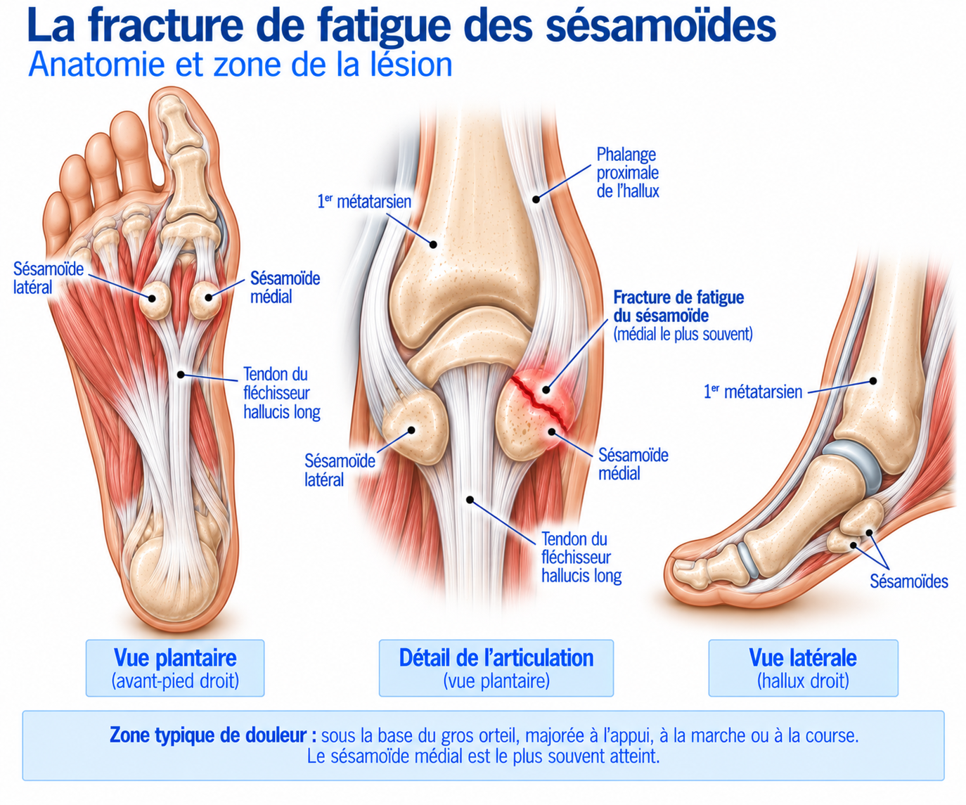
🔍 Click on the image to enlarge it
Why are the sesamoids under particular strain in runners?
During the terminal stance phase, the big toe gradually moves into dorsiflexion and body weight is transferred to the forefoot. The sesamoids are then literally caught between the head of the first metatarsal and the ground, and are subjected to considerable compression, shear and traction stresses, as well as to dorsiflexion of the first metatarsophalangeal joint.
These stresses become particularly high during fast running, sprints, accelerations, uphill running, jumping, dancing or gymnastics: activities involving repeated, forced dorsiflexion of the big toe are particularly associated with sesamoid stress fractures.
Epidemiology
Fracture of the sesamoids of the big toe is a relatively rare but significant injury in athletes, because the sesamoids are among the sites considered high-risk for stress fractures: the mechanical stresses on them are considerable and their blood supply is relatively poor, which exposes them to a risk of delayed union, or even non-union, without appropriate management.
It mainly affects athletes who place heavy demands on the forefoot in dorsiflexion: runners (fast running, sprints, accelerations, hills), jumpers, dancers and gymnasts. The medial sesamoid is generally the one most often affected, because its position directly beneath the head of the first metatarsal exposes it to greater stresses than the lateral sesamoid — a predominance also found in published case series.
Causes & risk factors
Several factors can contribute to a sesamoid stress fracture; however, no single characteristic should be considered the sole cause of the fracture.
Training factors
Sudden increase in mileage, return after a break, increase in the number of sessions, addition of interval sessions, more speed work or elevation gain, introduction of hills, abrupt change of surface, training with insufficient recovery.
Mechanical factors
Heavily loaded forefoot, marked dorsiflexion of the big toe, high load on the first ray, altered distribution of foot loading, certain foot morphologies.
Footwear-related factors
Footwear can greatly alter the stresses placed on the sesamoids: shoes that are very flexible in the forefoot or very minimalist, very thin soles, an abrupt change of shoes, shoes with a narrow toe box or that encourage marked flexion of the first ray. Conversely, in some athletes, a shoe with a more protective forefoot and a stiffer sole can reduce painful stresses.
Bone and biological factors
As with other stress fractures, factors that may reduce the bone’s capacity to withstand stress should be looked for: energy intake and energy availability, calcium and vitamin D intake, history of stress fractures, certain endocrine disorders, menstrual cycle disorders in female athletes, certain medications, significant recent weight loss. In an athlete with several stress fractures or recurrent stress fractures, investigating bone or metabolic factors is particularly relevant.
Symptoms
The main symptom is pain under the forefoot, at the base of the big toe; the patient can often point very precisely to the painful area. The pain is generally plantar and localised, located beneath the head of the first metatarsal, sometimes slightly medial or lateral.
A distinction must be made between a traumatic fracture, which occurs after an identifiable event (landing from a jump, fall, sudden hyperextension of the big toe, direct blow, crush injury) with sudden pain, and a stress fracture, which develops gradually: initial discomfort, then pain during exercise that appears earlier and earlier, possibly pain when walking and then, in advanced forms, pain at rest. The pain may appear at the start of a run, after a few kilometres, or during accelerations, hills, jumps and push-off. In early forms, a classic pitfall is that the pain disappears once exercise stops: improvement at rest does not mean that the bone has healed.
Progression may follow a stepwise pattern: pain only during strenuous exercise (initial stage), pain during normal running (intermediate stage), pain when walking (advanced stage), then pain at rest or persisting after activity (severe stage). A gradual decrease in the distance needed to trigger the pain should raise suspicion of a bone stress injury.
Clinical examination
The examination first looks for very localised pain. Direct pressure on the painful sesamoid usually reproduces the pain; the medial sesamoid, the lateral sesamoid, the head of the first metatarsal, the metatarsophalangeal joint, the plantar plate and the flexor hallucis longus tendon should each be examined separately.
Passive dorsiflexion of the first metatarsophalangeal joint may reproduce the pain, as forced dorsiflexion can compress the sesamoids beneath the metatarsal head. The examination should be performed weight-bearing when possible: walking, rising onto tiptoes, push-off, barefoot walking, possibly a jump if the situation allows — however, significant pain during push-off should prompt limiting provocative tests when a fracture is suspected.
Examination of the foot as a whole looks for hallux valgus, hallux rigidus, limited mobility of the first ray, plantar arch disorders, overpronation or supination, abnormal load distribution, calluses beneath the first metatarsal and any footwear issues, in order to understand why stresses are concentrated on the sesamoid.
Further tests
X-rays
First-line imaging: weight-bearing foot, AP, lateral and oblique views, with specific sesamoid views if necessary. Looks for fracture, displacement, fragmentation, first ray abnormality, osteoarthritis, hallux valgus, bipartite sesamoid. Caution: a normal X-ray does not rule out an early stress fracture — changes may appear late, and X-rays can be negative in a substantial proportion of early sesamoid stress fractures.
MRI
Particularly useful when the X-ray is normal or inconclusive. Can show bone oedema, a stress fracture, a bone stress reaction, soft tissue involvement, oedema around the sesamoid or associated plantar plate or tendon pathology. The investigation of choice to confirm an early stress fracture.
CT scan
Particularly valuable for analysing the fracture line, the cortex, displacement, fragmentation, healing or possible non-union. Useful for monitoring complex fractures; sagittal slices can be particularly informative when healing is in question.
Bone scintigraphy
Can show increased uptake in the sesamoid. Less widely used than MRI today, but may still be useful in some complex diagnostic situations when MRI is not available.
Differential diagnoses
Bipartite sesamoid
A common anatomical variant, often asymptomatic, in which the sesamoid consists of fragments separated by a synchondrosis. A fracture line is generally more irregular, sharper and lacks a regular cortical margin, whereas a bipartite sesamoid has smoother contours and corticated edges; comparison with the opposite side can help, but the distinction may remain difficult on a standard X-ray.
Sesamoiditis
Painful irritation of the sesamoid complex without a true break in the bone's structure — the two conditions can cause very similar symptoms.
Sesamoid non-union
Pain persisting several months after the fracture, no improvement despite appropriate offloading, persistent fracture or fragmentation on imaging, mobility at the fracture site, pain consistently returning with loading.
Joint conditions
Metatarsophalangeal osteoarthritis, hallux rigidus, synovitis, gout — the pain is more articular and less strictly focal over the sesamoid.
Tendon conditions
Flexor hallucis longus tendinopathy, flexor hallucis brevis injury — pain is often reproduced by stretching or resisted contraction of the tendon rather than by bone palpation alone.
Other
Plantar plate injury, metatarsalgia, capsuloligamentous injury, foreign body, fracture of the head of the first metatarsal.
Classification
Grade 1
Stress reaction: pain on exertion, normal X-ray, MRI showing bone oedema without a complete fracture.
Grade 2
Non-displaced stress fracture: fracture visible on MRI, CT or X-ray, without displacement, stability preserved.
Grade 3
Complex fracture: displacement, fragmentation, unstable complete fracture, significant involvement of the sesamoid complex.
Grade 4
Chronic course: non-union, persistent pain, chronic fragmentation, failure to heal despite appropriate treatment.
Treatment
Phase 1 – Bone protection and offloading
When a stress fracture is diagnosed, the priority is to remove the stresses that are maintaining the injury: running, sprinting, jumping, dancing and sports involving strong push-off from the big toe generally need to be stopped temporarily. The level of offloading depends on the pain and the severity of the fracture. Depending on the case, a rigid-soled shoe, a forefoot offloading shoe, a post-operative shoe, a walking boot, or even stricter immobilisation in some forms may be used — because sesamoid stress fractures are considered high-risk fractures, some forms require more offloading or greater weight-bearing restriction than low-risk stress fractures.
There is no single timeframe: a period of several weeks is generally needed, with protocols of 4 to 8 weeks of restricted weight-bearing reported in the literature in particular, followed by a gradual return to weight-bearing. The duration must, however, be adapted to the pain, imaging findings, healing and type of fracture.
Orthoses play an important role in reducing the pressure exerted directly on the sesamoid: U-shaped offloading, a cut-out beneath the sesamoid, an insole adapted to the first ray, a correctly positioned metatarsal pad (a poorly positioned pad can actually increase pressure — the insole must therefore be individualised). Footwear is modified temporarily, favouring a sufficiently stiff sole, good support, enough room for the toes and limited painful flexion of the first ray. Painkillers and anti-inflammatories may be used if necessary, but with caution: above all, they must not be used to mask the pain in order to return to running prematurely, as pain is a useful signal for adjusting load.
Phase 2 – Return to weight-bearing and rehabilitation
When the pain has decreased sufficiently, weight-bearing is gradually increased: from offloading to protected weight-bearing, then normal weight-bearing, then functional exercises — progression depends on the pain and on healing.
Rehabilitation often begins with impact-free work: ankle mobility, toe mobility within pain-free ranges, triceps surae strengthening, strengthening of the intrinsic foot muscles, balance, proprioception, lower limb control.
Foot and calf strengthening
The foot must gradually regain its capacity to withstand stress. Possible exercises: short foot (activation of the plantar arch, 3 × 10 to 15 repetitions), toe flexion (progressive work of the intrinsic muscles), calf raises (first double-leg, then single-leg), single-leg balance (progressing to more demanding surfaces).
The triceps surae plays a major part in push-off: progression can be made in levels — double-leg calf raises, then single-leg raises, then long sets for endurance, then plyometric work. Progression should be carried out without significant sesamoid pain.
Reintroducing impacts and returning to running
Once normal walking and strengthening exercises are pain-free, double-leg jumps, jumps on the spot, single-leg hops, bounds and then easy running can be gradually reintroduced. Progression must be slow, as the sesamoid is subjected to considerable stress during push-off.
The return to running ideally follows a stepwise progression: pain-free brisk walking, alternating running/walking (1 to 2 min of running for 1 min of walking), easy continuous running, increased duration, increased mileage, reintroduction of hills, reintroduction of accelerations, then a return to interval training and specific sessions.
Before returning, the ideal is no pain when walking or on palpation, satisfactory big toe mobility, pain-free calf raises, tolerated single-leg exercises, pain-free progressive jumps and no delayed pain after exercise. In a high-risk stress fracture, the return should also take radiological healing into account when necessary.
Complementary treatments
Sesamoid offloading orthoses
U-shaped offloading, cut-out beneath the sesamoid, individualised metatarsal pad: these reduce the pressure exerted directly on the sesamoid. A poorly positioned insole can actually make the pain worse.
Suitable footwear
Sufficiently stiff sole, good support, enough room for the toes, limited painful flexion of the first ray. Can reduce dorsiflexion of the big toe during push-off.
Painkillers and anti-inflammatories
May be used if necessary, but with caution in a stress fracture: they must not mask the pain to the point of allowing a premature return to running.
Addressing bone and biological factors
Energy intake and energy availability, calcium and vitamin D intake, screening for endocrine disorders or menstrual cycle disorders in female athletes in the event of recurrent stress fractures.
Surgery
Surgery may be considered in cases of displaced or unstable fracture, symptomatic non-union, persistent pain despite properly conducted conservative treatment, significant fragmentation, or inability to return to sport despite several months of treatment. There are two main options: internal fixation of the fracture and sesamoidectomy, total or partial — the choice depends in particular on the anatomy and on whether the sesamoid can be preserved.
When the sesamoid can be preserved, osteosynthesis (screw, fixation adapted to the fragment, possibly bone grafting in cases of non-union) preserves its biomechanical role; a systematic review suggests that internal fixation offers the best chance of returning to the previous level of sport, although the available data remain limited and heterogeneous.
Sesamoidectomy involves removing all or part of the sesamoid. It may be considered when the sesamoid is highly fragmented, in cases of painful non-union, when bone preservation is not possible, or after failure of conservative treatment. It often provides significant relief, but it is not a trivial procedure: because the sesamoids play an important biomechanical role, their removal can lead to weakness of hallux flexion, altered push-off, residual pain, a painful scar, a neuroma, hallux valgus or hallux varus. The risk depends in particular on which sesamoid is removed and on the quality of the repair of the capsulotendinous apparatus.
To simplify: osteosynthesis preserves the sesamoid and the biomechanics but involves more technical surgery and requires bone healing; sesamoidectomy removes the painful focus, with recovery that is sometimes faster, at the cost of a risk of altered biomechanics and residual plantar pain. Overall, the literature suggests that sesamoidectomy may allow a faster return, whereas internal fixation offers a better prospect of returning to the previous level of sport. The time to return to sport then depends on the type of procedure, healing, the sport, the level of competition and muscle recovery, and should be based on pain, healing, strength, mobility, push-off capacity and functional tests.
Recovery time
A distinction should be made between clinical healing, radiological healing, functional recovery and return to the previous level of sport: these four stages do not always occur at the same time, and the literature shows that recovery can be lengthy. In a series of young athletes with sesamoid fractures, around 85% were able to return to sport, but the mean time between diagnosis (or the start of treatment) and being pain-free with clearance to return was around 161 days, i.e. more than five months — however, this was a young population and a retrospective series: this timeframe should not be regarded as a mandatory duration.
Several factors explain this sometimes prolonged course: considerable mechanical stresses, the thin layer of tissue between the bone and the ground, repeated compressive stresses, a relatively poor blood supply, the difficulty of fully offloading the first ray, daily loading during walking, and the risk of returning to sport too early. These are among the reasons why sesamoid stress fractures are considered high-risk fractures.
Prevention
Preventing recurrence relies on several areas. Load management: avoid sudden increases in mileage, an overly rapid return and an accumulation of speed sessions. Footwear: choose a shoe suited to the foot and to the level of load, avoiding models that are too flexible or minimalist in the forefoot if these cause pain.
Strengthening should be maintained over the long term: calves, intrinsic foot muscles and lower limb muscles. Finally, when changing shoes or running technique, it is better to change only one variable at a time, gradually, rather than combining several simultaneous changes.
Frequently asked questions
Can you run with a sesamoid fracture?
Is a sesamoid fracture serious?
Can a stress fracture be invisible on an X-ray?
What is the difference between sesamoiditis and a fracture?
Can you have a fracture without trauma?
Is a bipartite sesamoid an old fracture?
How long does it take to heal?
Is a CT scan necessary?
Do I need an MRI?
Is surgery mandatory?
Can you return to sport after a sesamoidectomy?
Can you keep cycling or swimming during treatment?
Common mistakes
Carrying on running "as long as it's bearable": a stress fracture is not just a tendinopathy, and continuing to load the painful bone can delay healing and lead to worsening.
Being reassured by a normal X-ray: a normal early X-ray does not rule out a stress fracture.
Confusing a fracture with a bipartite sesamoid: a separation line may correspond to an anatomical variant or to a true fracture.
Returning as soon as the pain disappears: the pain sometimes disappears before the bone has fully recovered its mechanical capacity.
Relying solely on painkillers, when pain should above all lead to adjusting the load.
Neglecting footwear, even though it can greatly alter the stresses placed on the sesamoids.
Going straight back to interval training: accelerations and hills place considerable stress on the first ray.
Key points — Institut Basque du Coureur
Fracture of the sesamoids of the big toe is a relatively rare but significant injury in athletes, whether traumatic or, very often in runners, a stress fracture. The medial sesamoid is the one most often affected. The typical pain is plantar, beneath the head of the first metatarsal, at the base of the big toe, and is often reproduced by palpation and by dorsiflexion of the big toe. Diagnosis is based on the history, clinical examination, X-rays, MRI when the X-ray is normal or inconclusive, and CT for certain fractures or to assess healing — a normal X-ray therefore does not rule out a stress fracture. Initial treatment generally involves stopping impact activities, appropriate offloading or immobilisation, protective footwear, possibly an offloading orthosis, a gradual return to weight-bearing, rehabilitation and then the gradual reintroduction of impacts and running. These fractures are considered high-risk and should be taken more seriously than simple mechanical forefoot pain; surgery is reserved for selected situations (displacement, instability, non-union, significant fragmentation, failure of conservative treatment), with the choice between preservation/fixation of the sesamoid and sesamoidectomy taking into account the level of sport and the biomechanics of the first ray. In runners, persistent, very localised pain beneath the big toe should suggest a stress fracture, even in the absence of trauma and even if the first X-ray is normal.
- McInnis KC, Ramey LN. High-Risk Stress Fractures: Diagnosis and Management. PM R. 2016;8(3 Suppl):S113-S124.
- Robertson GAJ, Goffin JS, Wood AM. Return to sport following stress fractures of the great toe sesamoids: a systematic review. Br Med Bull. 2017;122(1):135-149.
- Boden BP, Osbahr DC, Jimenez C. Low-risk and high-risk stress fractures: evaluation and management. J Am Acad Orthop Surg. 2001;9(2):80-90.
- Robertson GAJ, Wood AM. Return to sport following stress fractures of the great toe sesamoids: a systematic review. Br Med Bull. 2017;122(1):135-149.
- Patel S, Roth M, et al. Stress fractures of the foot and ankle in athletes. Sports Med Arthrosc Rev. 2014;22(3):160-170.
- Stein CJ, Sugimoto D, Slick NR, et al. Hallux sesamoid fractures in young athletes. Clin J Sport Med. 2019;29(6):441-447.
- Saxena A, Krisdakumtorn T. Return to activity after sesamoidectomy in athletically active individuals. Foot Ankle Int. 2003;24(5):415-419.
- Lee S, et al. Sesamoidectomy in Athletes: Outcomes From 2-Centers. Foot Ankle Orthop. 2021.
- Lim S, Liem B. First Metatarsophalangeal Joint Pain in Athletes: Diagnosis, Management, and Return to Play Considerations. Curr Sports Med Rep. 2023;22(6):217-223.
Similar injuries
Plantar fasciopathy
Heel pain caused by irritation of the plantar fascia, very common in runners.
Achilles tendinopathy
Inflammation of the Achilles tendon causing pain and stiffness at the heel, the most common injury in runners.
Blisters in runners
Fluid-filled pockets under the skin caused by friction, heat and moisture, common in running, trail running and marathons.