
Definition
Pes anserinus tendinopathy refers to pain located on the anteromedial aspect of the knee, at the common insertion of several tendons on the inner surface of the tibia. The “pes anserinus” (literally “goose’s foot”) essentially corresponds to the junction of the tendons of three muscles: the sartorius, the gracilis and the semitendinosus.
In the immediate vicinity of this insertion lies the pes anserine bursa, a small structure that reduces friction between the different tissues. This is why the term pes anserine pain syndrome is often more accurate than “pes anserinus tendinopathy”: the pain may in fact come from the tendons themselves, from the bursa, from the interface between these structures, or more broadly from the medial tissues of the knee.
Distinguishing between bursitis and tendinopathy is sometimes clinically difficult, as the two structures are anatomically extremely close.
Anatomy
The pes anserinus is formed mainly by the tendons of three muscles that converge on the anteromedial aspect of the proximal tibia. The sartorius, a superficial muscle, contributes to hip flexion, abduction and external rotation as well as knee flexion. The gracilis, a medial thigh muscle, contributes to hip adduction, knee flexion and medial rotation of the leg. The semitendinosus, one of the hamstrings, contributes to hip extension, knee flexion and medial rotation of the leg.
The actual anatomy is, however, more variable than a simplified representation would suggest: recent anatomical studies show numerous variations in the configuration and tendon expansions of the pes anserinus.
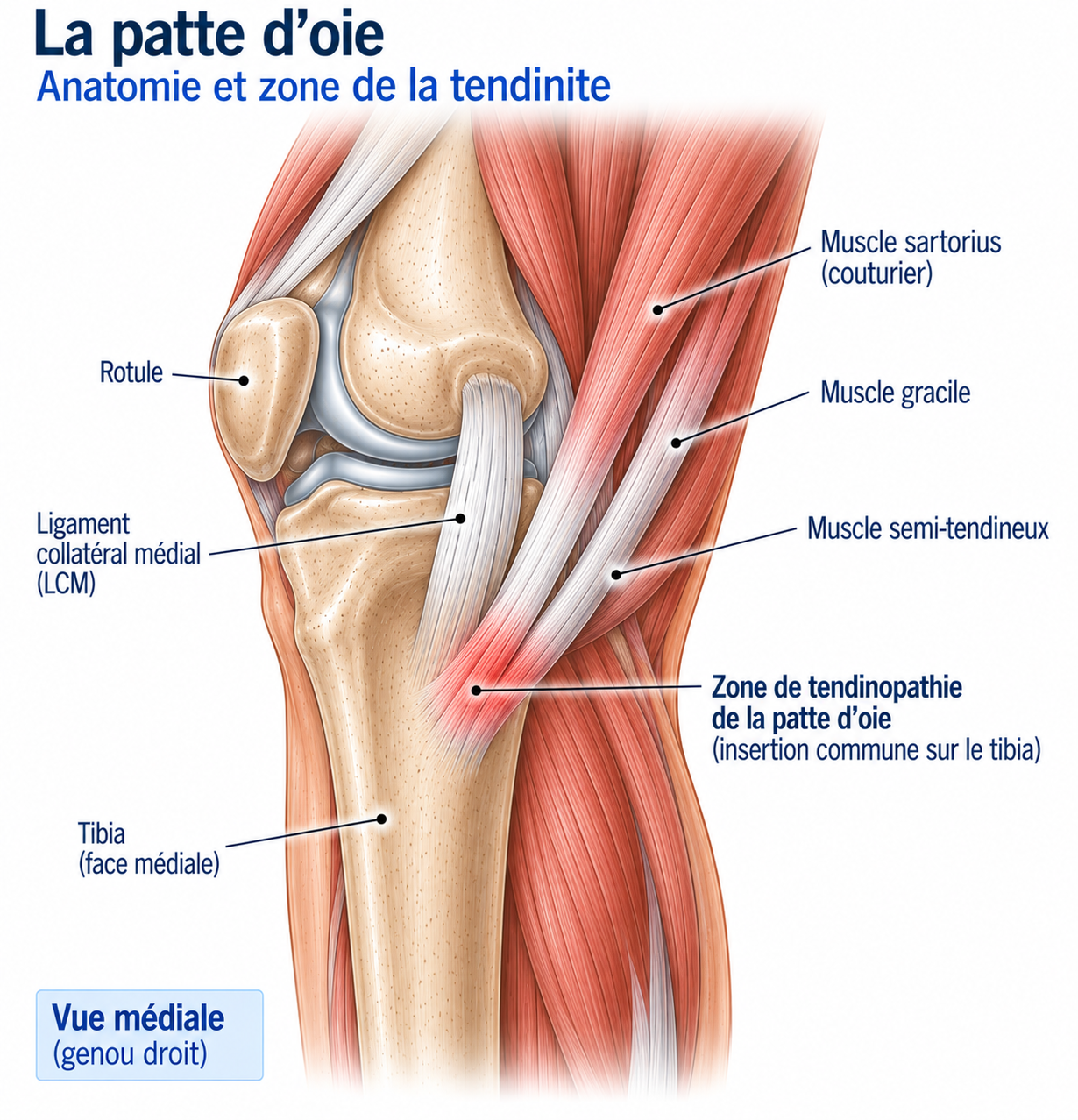
🔍 Click on the image to enlarge it
The pes anserine bursa
Beneath the pes anserinus tendons lies a synovial bursa, the pes anserine bursa, whose role is to help the different structures glide over one another. It can become painful and inflamed in certain situations — this is then referred to as anserine bursitis. This distinction is important because pain at the pes anserinus does not automatically mean that the tendon is diseased.
Function and biomechanics
The three pes anserinus muscles have different but complementary functions: they contribute to knee flexion, medial rotation of the leg, dynamic stabilisation of the knee, lower limb control and stabilisation of the pelvis and hip. The tendinous and fascial structures of this region form a genuine stabilising complex for the medial aspect of the knee.
The pes anserinus is loaded during movements that combine knee flexion, medial rotation, knee stabilisation and hip control. Stresses increase in particular during running, uphill sections, stairs, changes of direction, lateral movements, single-leg exercises and repeated knee flexion movements; overload may affect the tendons directly or increase the stresses placed on the bursa.
Epidemiology
Pes anserine pain syndrome affects both active and non-active people: it can also appear in a context of everyday mechanical overload or associated knee disease. It is frequently found in association with knee osteoarthritis, excess weight, diabetes or older age, although these factors should not be interpreted as sole causes.
Causes & risk factors
As with other tendinopathies, there is usually an imbalance between the load applied and the capacity of the tissues to withstand it. This can appear after an increase in mileage, a return to sport, an increase in elevation gain or stairs, a change in running technique, an increase in strength sessions or repeated changes of direction — often gradually, without any specific trauma.
Tendinopathy or bursitis?
In tendinopathy, the pain comes mainly from the tendon structures and is usually triggered by stretching or contracting the muscles involved. In bursitis, the pain comes mainly from the bursa and may be particularly sensitive to local pressure, to contact with certain surfaces or during certain repeated movements. In practice, the two phenomena may coexist — hence the preference for the term “pes anserine pain syndrome” — and conservative treatments are in any case largely similar in both situations.
Contributing factors
Load-related factors: sudden increase in mileage, return to running, increase in elevation gain, more hill sessions, more stairs, more strength sessions.
Biomechanical factors: poor lower limb control, hip muscle weakness, hamstring strength deficit, poor single-leg control, increased medial knee stress.
Associated factors: knee osteoarthritis, excess weight, diabetes, older age — although these should not be interpreted as sole causes.
Symptoms
The patient often describes pain on the inner side of the knee, a few centimetres below the joint, that has come on gradually. It is often triggered by running, stairs, uphill sections, squats or single-leg movements; in some cases, it is also present at rest or at night — although significant or atypical night pain should prompt a search for another cause.
The pain is usually medial and slightly below the joint line, often located a few centimetres below the medial joint line of the knee — a location that helps distinguish it from meniscal, ligament or patellofemoral pain, although symptoms may overlap. In runners, the pain may appear during the run, after the run, the next day, or on uphill or downhill sections: a recent change in volume or elevation gain should always be looked for. Stairs can also reproduce the pain, particularly going up, going down or after several flights.
Clinical examination
The examination should establish the precise location of the pain, tenderness on palpation and pain on contraction of the pes anserinus muscles, knee mobility, the strength of the quadriceps, hamstrings and hip muscles, lower limb control, as well as meniscal, ligament and neurological signs: the diagnosis should not rest solely on a tender spot.
Palpation looks for pain specifically at the pes anserinus insertion, comparing with the opposite side. However, the diagnostic accuracy of the clinical examination alone is imperfect: a study comparing clinical diagnosis with MRI findings found relatively low sensitivity and specificity. Contraction tests targeting the semitendinosus, gracilis and sartorius (resisted knee flexion, sometimes combined with internal rotation of the leg) may aim to reproduce the usual pain.
Examination of the hip looks for abductor weakness, poor pelvic control, pain or limited mobility — a proximal deficit can alter the stresses placed on the knee during running or single-leg movements. The squat, particularly the single-leg squat, makes it possible to assess pain, knee stability, lower limb alignment, hip control and tolerance to flexion. In runners, gait analysis mainly looks for recent changes in load (volume, speed, elevation gain, shoes, hills), which are often more decisive than looking for a biomechanical abnormality in isolation.
Further tests
Ultrasound
Allows assessment of the pes anserinus tendons, the pes anserine bursa, the surrounding tissues, certain tendon lesions and any fluid collection, with the possibility of comparison with the opposite side.
MRI
Can look for anserine bursitis, tendon abnormalities, soft tissue oedema, osteoarthritis, a meniscal or ligament injury, a stress fracture or other knee conditions. Particularly useful when the medial pain does not fully match the classic picture.
Differential diagnoses
Medial meniscal tear
To be considered in the presence of joint line pain, locking, catching or pain on rotational movements.
Medial osteoarthritis
Pain usually closer to the joint line, which may be accompanied by stiffness, limited mobility or crepitus.
Medial collateral ligament injury
To be considered after a valgus injury or in the presence of ligament pain.
Semimembranosus tendinopathy
May cause posteromedial knee pain.
Semitendinosus tendinopathy
May be located closer to the pes anserinus.
Patellofemoral pain syndrome
Pain usually more anterior.
Popliteal cyst
Pain usually more posterior.
Stress fracture
To be considered in the case of persistent impact-related pain, particularly after a sudden increase in load.
Classification
Stage 1
Pain only after activity.
Stage 2
Pain during activity but still allowing sport to be practised.
Stage 3
Pain limiting running, stairs or training.
Stage 4
Pain present at rest or during daily activities.
Treatment
Load management and reducing irritability
Treatment is based on load management, progressive strengthening, correction of contributing factors and a gradual return to sport. The first step is to identify the activities that trigger symptoms (recent mileage, hills, stairs, interval training, changes of direction, single-leg work, hamstring strengthening): it is often possible to keep running while temporarily reducing volume, speed, elevation gain, hills and intervals — a slow, easy run on flat ground may be well tolerated whereas a hill session triggers symptoms. The knee's response during activity and over the following 24 hours should guide progression.
As irritability decreases, the goals are to reduce pain, maintain mobility, stay physically active and begin progressive reloading: hamstring isometrics (glute bridge held for 20 to 40 seconds, 3 to 5 repetitions) and quadriceps isometrics (resisted knee extension, 30 to 45 seconds, 4 to 5 repetitions), staying within an acceptable tolerance zone.
Progressive strengthening of the medial chain
Strengthening of the hamstrings (including the semitendinosus) is introduced gradually: glute bridge, single-leg bridge, leg curl, hip thrust, Romanian deadlift, starting with moderate loads and then increasing them progressively. Strengthening of the gracilis and of medial control involves hip adduction, an adapted Copenhagen plank, knee flexion exercises and single-leg work — the aim being not to isolate the gracilis perfectly but to strengthen the entire medial chain.
Strengthening of the sartorius and the hip favours functional work (step-ups, stair climbing, lunges, single-leg squats, hip control exercises) rather than seeking absolute isolation. The quadriceps should also be strengthened (squats, leg press, knee extension, split squats, step-ups): a sufficiently strong quadriceps helps distribute stresses better across the entire lower limb.
Unilateral work and the kinetic chain
Unilateral work gradually brings rehabilitation closer to the demands of sport: single-leg squats, step-downs, lunges, single-leg Romanian deadlifts, bench step-ups — while monitoring pain, stability, pelvic control and knee alignment.
Rehabilitation should include the entire kinetic chain: quadriceps, hamstrings, glutes, abductors, adductors, calves and trunk muscles, in order to restore overall lower limb function — the pes anserinus is not a structure that works in isolation. Mobility should be assessed, but there is no need to systematically aim for great flexibility; stretching can be used when a deficit has been identified, but it is not the main treatment, and aggressive stretching of a painful area should be avoided.
Gradual return to running and sport
In runners, progression may follow this sequence: brisk walking, slow easy running, continuous running, gradual increase in volume, strides, interval training, hills, then a return to the normal programme — elevation gain often needs to be reintroduced later than running on flat ground. For example, a runner might temporarily replace a hilly 10 km with intervals by 30 to 40 minutes on flat ground at low intensity, then progress in duration, volume, pace, strides, hills and intervals.
For team sports, lateral movements, braking, changes of direction, accelerations and decelerations also need to be reintroduced gradually. Before a full return, the criteria are low or no pain, no significant pain in daily activities, good tolerance of running and unilateral exercises, satisfactory lower limb strength and stability, and no worsening within 24 hours.
Complementary treatments
Taping
May sometimes temporarily reduce pain, improve comfort or make certain exercises easier, but does not directly treat the cause of the tendinopathy.
Massage and manual therapy
May provide symptomatic relief and temporarily improve mobility, but does not replace active rehabilitation.
Shockwave therapy
Limited specific data; may be considered in some chronic forms but is not a first-line treatment.
Corticosteroid injection
May sometimes be considered for clearly identified anserine bursitis that is very symptomatic and resistant to conservative treatment — but should not be performed simply on the basis of medial knee pain, as the diagnostic accuracy for this syndrome is imperfect.
PRP (platelet-rich plasma)
Insufficient data to make it a standard treatment; possibly considered in certain specific chronic situations, after properly conducted active management.
Surgery
Surgery is very rarely necessary. It may be considered only in very specific situations, after diagnostic confirmation, prolonged conservative treatment and failure of properly conducted rehabilitation. Most patients should be managed without surgery.
Recovery time
Recovery time varies: a mild form may improve within a few weeks, whereas a chronic form may require several months. It depends in particular on how long the symptoms have been present, the sporting load, the presence of associated bursitis, the existence of osteoarthritis, the quality of rehabilitation and the ability to adjust loads. Persistent pain does not necessarily mean that the tendon is severely damaged.
Prevention
Preventing recurrence relies mainly on a reasonable progression of load, regular strengthening, unilateral work, strengthening of the hip, hamstrings and quadriceps, and a gradual return to hills and changes of direction. Sudden changes in volume or elevation gain should be particularly avoided.
Frequently asked questions
Where exactly is pes anserinus pain located?
Is it really a tendinopathy?
Can I run?
Can I cycle?
Do I need an MRI?
Are injections effective?
Is shockwave therapy necessary?
Is it linked to osteoarthritis?
Can you develop pes anserinus pain without playing sport?
Common mistakes
Assuming that any medial knee pain is pes anserinus pain, when this pain has many possible causes.
Giving an injection without confirming the diagnosis, when the accuracy of clinical diagnosis alone is limited.
Stopping activity completely, when complete rest is generally not necessary.
Going straight back to hill sessions, which can represent a significant increase in load.
Working only on the painful area, without including the hip, quadriceps and hamstrings in rehabilitation.
Focusing solely on flexibility, when the capacity to tolerate load is more important.
Going straight back to changes of direction, which should be reintroduced gradually in athletes.
Key points — Institut Basque du Coureur
"Pes anserinus tendinopathy" is in fact a pain syndrome of the pes anserinus region, which may involve the tendons of the sartorius, gracilis and semitendinosus, but also the pes anserine bursa. The classic picture is pain on the inner side of the knee, slightly below the joint line. Diagnosis should nevertheless be cautious, as many other conditions can cause medial knee pain. Management relies mainly on load management, progressive strengthening, work on the hip and lower limb, and a gradual return to running and sport. In runners, mileage, speed, elevation gain, hills and changes of direction should be monitored in particular. The aim is not simply to make the pain go away: the goal is to gradually make the knee able to withstand the real demands of sport again. Pain at the pes anserinus is not always a tendinopathy: before treating, you need to be sure what you are treating.
- Curtis BR, Huang BK, Pathria MN, Resnick DL, Smitaman E. Pes Anserinus: Anatomy and Pathology of Native and Harvested Tendons. AJR Am J Roentgenol. 2019;213(5):1107-1116.
- Levent A, et al. Pes Anserine Bursitis. StatPearls.
- Şahin N, et al. Poor Accuracy of Clinical Diagnosis in Pes Anserine Tendinitis Bursitis Syndrome. Indian J Orthop. 2022.
- Klauser AS, Tagliafico A, Allen GM, et al. Clinical and sonographic evaluation of the pes anserinus region.
- Kim YC, et al. Pes anserinus and anserine bursa: anatomical study. Anat Cell Biol. 2014;47(2):127-131.
- Çavuş F, Erbek E, Bolatlı G. A systematic review of pes anserinus variations, their anatomical types, prevalence, and clinical reflections. Surg Radiol Anat. 2025;48(1):29.
- Możdżeń K, et al. Morphometry and functional topography of the pes anserinus complex: A standardized cadaveric study. Ann Anat. 2026.
- Pes anserine bursitis: incidence in symptomatic knees and clinical presentation. Eur Radiol. 2005.
- Knee Bursae: A Comprehensive Review of Clinical Evaluation, Imaging Differentiation, and the Expanding Role of Biologic Therapies. 2025.
Similar injuries
Iliotibial band syndrome
Irritation of the iliotibial band on the outer side of the knee, very common in runners.
Patellofemoral pain syndrome
Anterior knee pain caused by poor patellar tracking, accounting for 25 to 40% of knee injuries in runners.
Meniscal injury in runners
Damage to the knee meniscus through a tear or progressive degeneration, common in runners over 35.