
Definition
Patellofemoral pain syndrome, now more often referred to as patellofemoral pain (PFP), is pain located around or behind the kneecap, usually triggered by activities that increase the stresses on the patellofemoral joint: running, going down and up stairs, squats, lunges, jumping, prolonged sitting with the knee bent, or cycling in certain situations.
The pain is usually located around the kneecap, behind it or along its edges; it is sometimes harder to pinpoint precisely.
It is important to understand that patellofemoral pain syndrome is not simply a problem of a “badly positioned” kneecap. It is a multifactorial condition involving the load applied to the knee, muscle capacity, quadriceps strength, the hip muscles, lower limb biomechanics, movement technique, activity level, as well as psychological and contextual factors. Recent international guidelines therefore favour an individualised approach rather than the systematic correction of a supposed biomechanical abnormality.
Anatomy
The patellofemoral joint is the joint between the kneecap (patella) and the femoral trochlea. The patella is a sesamoid bone located within the quadriceps tendon; it improves the quadriceps’ lever arm, transmits forces between the quadriceps and the patellar tendon, protects the front of the knee and helps guide the flexion-extension movement.
The back of the patella is covered with articular cartilage, which is particularly thick because patellofemoral stresses can become very high during squats, on stairs, when running or when jumping.
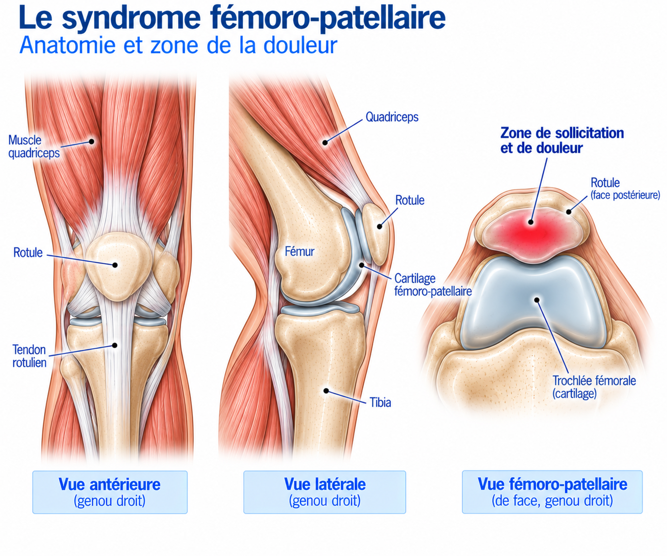
🔍 Click on the image to enlarge it
Patellofemoral biomechanics
As the knee bends, the patella gradually engages in the trochlea. The force exerted by the quadriceps increases the compressive stresses between the patella and the femoral trochlea: the greater the load, the greater the patellofemoral force. This is why certain activities are particularly symptomatic: deep squats, lunges, going down stairs, downhill running, jumping and landing.
But pain is not simply proportional to the pressure exerted on the joint. A knee can withstand considerable stress without pain, and conversely a lightly loaded knee can become painful. Pain also depends on the tissues’ capacity to tolerate load, recent exposure to exercise, recovery, nervous system sensitivity, the psychological context and previous experience of pain.
Epidemiology
Patellofemoral pain is one of the most common causes of anterior knee pain. It particularly affects adolescents, young adults, athletes, runners and people who play sports involving jumping and changes of direction. It is especially common in runners.
In some patients, it can become chronic, with a prolonged course despite initial management. This is why it is important not to limit treatment to short-term pain relief, but also to gradually restore the knee’s capacity to tolerate load.
Causes & risk factors
There is no single factor responsible for patellofemoral pain: it is a multifactorial condition combining elements related to load, muscle capacity, biomechanics and the patient’s individual profile.
The role of the quadriceps
The quadriceps is essential to patellofemoral function: it extends the knee, absorbs loads, controls knee flexion during running and dynamically stabilises the lower limb. Weakness or loss of capacity in the quadriceps can reduce the knee’s tolerance to stress. However, a classic mistake must be avoided: patellofemoral pain is not simply due to a “too weak” quadriceps. Strengthening it is extremely important, but it must be part of a comprehensive management plan.
The role of the hip
The hip muscles — gluteus medius, gluteus maximus, external rotators, abductors — help control the lower limb, particularly hip adduction, internal rotation and dynamic knee alignment. Poor control of the lower limb during single-leg stance can increase certain patellofemoral stresses. But here again, increased knee adduction or rotation is not on its own the cause of the pain: it is one factor among others.
Load-related factors
Increased mileage, returning to running, a sudden increase in intensity or elevation gain, introducing interval training, returning to a sport after a break, an increase in the number of sessions.
Muscular factors
Quadriceps strength deficit, hip strength deficit, overall muscle capacity deficit, muscle fatigue.
Biomechanical factors
Insufficient control of the lower limb, changes in cadence, running technique, reduced ankle or hip mobility.
Individual factors
History of patellofemoral pain, activity level, sleep, recovery, stress, fear of movement.
The typical runner scenario
The classic picture is that of a runner who increases their training — for example more mileage, added interval sessions and significant elevation gain. The knee then gradually becomes painful: at first only after running, then during running, then on stairs or when sitting for long periods. This is often a sign that the knee’s current capacity is being exceeded by the load imposed on it.
Symptoms
The main symptom is anterior knee pain, described as pain around or behind the kneecap, pressure, discomfort, burning or a feeling of an “irritated” knee. It may be present during or after running, when going down stairs, during squats or lunges, or after prolonged sitting.
The well-known “movie sign” (or “cinema sign”) refers to pain that appears after sitting for a long time with the knees bent. The patient typically explains: “After 30 minutes sitting down, I need to straighten my leg because my knee starts to hurt.” This position increases the duration of patellofemoral compression, and this sign is frequently found in patients with PFP.
Clinical examination
The diagnosis is essentially clinical. History-taking looks at the exact location of the pain, how it started, how it has progressed, aggravating and relieving factors, any history of trauma, and any episodes of instability, swelling or locking.
The functional examination then relies on several tests that reproduce the pain: the squat (pain, depth, knee control, pelvic stability, movement quality), the single-leg squat (lower limb control, hip adduction, pelvic stability), the step-down (stepping down from a step, knee and pelvic control) and the lunge (load tolerance, lower limb control, quadriceps and hip strength). An assessment of the strength of the quadriceps, hamstrings, gluteus medius and maximus and calf completes the examination; ideally, this strength can be measured objectively using a dynamometer, isokinetic tests, force platforms or standardised functional tests, in order to track progress rather than relying solely on the patient’s own perception.
Further tests
X-ray
Generally not essential as a first-line investigation for typical patellofemoral pain. It may be requested in the case of atypical symptoms, persistent pain despite appropriate treatment, trauma, suspected osteoarthritis or to look for a bone abnormality.
MRI
Useful when a significant cartilage lesion, meniscal or ligament injury, instability, stress fracture or other intra-articular condition is suspected. Note: MRI very frequently shows abnormalities (irregular cartilage, hyperintense signals, oedema, small meniscal lesions) that are not necessarily responsible for the pain — the diagnosis must always remain clinical.
Differential diagnoses
Patellar tendinopathy
Pain located more at the lower pole of the kneecap or on the patellar tendon, typically triggered by jumping and explosive contractions.
Quadriceps tendinopathy
Pain located at the upper pole of the kneecap.
Iliotibial band syndrome
Lateral knee pain, particularly common in runners.
Meniscal tear
More localised pain, sometimes associated with locking, catching or pain during twisting movements.
Patellofemoral osteoarthritis
More common with age; pain when going up or down stairs, crepitus, stiffness, gradual loss of function.
Patellar instability
To be considered in the presence of a feeling of giving way, dislocation, subluxation or apprehension.
Cartilage lesion
May cause patellofemoral pain but should not automatically be confused with classic PFP.
Hoffa's syndrome
Anterior/infrapatellar pain, sometimes promoted by knee hyperextension.
Referred pain
Possible origin in the hip, lumbar spine or a peripheral nerve — to be systematically investigated.
Classification
Stage 1 — mild pain
Pain after activity, little limitation, sport practised almost normally.
Stage 2 — moderate pain
Pain during certain activities, stairs or squats difficult, reduced training volume.
Stage 3 — severe pain
Pain during daily activities, significant limitation of sport, pain in prolonged positions.
Stage 4 — persistent form
Chronic symptoms, severe limitation, possible psychological or occupational impact, failure of several previous treatments.
Treatment
Education and treatment principles
The treatment of patellofemoral pain rests on two pillars: education and progressive exercise. The most recent evidence confirms that targeted knee strengthening, with or without hip work depending on the patient's profile, forms the foundation of management. The aim is not to "put the kneecap back in place", but to reduce pain, increase muscle capacity, improve load tolerance, restore function and enable a return to sport.
Load management
Load management is often the first step in treatment. It is generally not necessary to stop sport completely: mileage, speed, elevation gain, session frequency, squat depth or jumping volume can be temporarily adjusted. For example, a runner whose pain is triggered after 8 km may temporarily run 4 to 5 km without significant pain, then gradually increase. The aim is to stay active while giving the knee sufficient capacity to recover.
Mild to moderate pain during activity may remain acceptable if it stays under control, does not gradually increase during the session, does not cause any major change in technique, returns to its usual level within the following hours and the knee is not significantly more painful the next morning. The principle is monitoring the response to load rather than systematically forbidding any pain.
Rehabilitation — phase 1: reduce pain and restore movement
The aims of this first phase are to reduce irritability, maintain tolerated activities, restore mobility and begin strengthening. Possible exercises include isometric quadriceps contractions, terminal knee extension, mini-squats, bridges and hip exercises. Intensity is gradually increased according to tolerance.
Rehabilitation — phase 2: quadriceps strengthening
The quadriceps is a key target for strengthening. Progression can follow a sequence such as: wall sit → partial squat → squat → loaded squat → single-leg squat → dynamic exercises. Exercises may include the squat, leg press, knee extension, step-up, split squat and lunge. The aim is to gradually restore the knee's capacity to withstand stress.
Rehabilitation — phase 3: hip strengthening
Depending on the patient's profile, hip work is added: hip abduction, single-leg bridge, hip thrust, step-down, lunge, single-leg squat, abductor and rotator work. Combined hip + knee work is particularly recommended by international guidelines.
Rehabilitation — phase 4: functional work and return to sport
Once strength is sufficient, work continues with deeper squats, lunges, step-downs, stair climbing, single-leg work and dynamic exercises, followed by jumps, landings, changes of direction, accelerations and decelerations.
For runners, running is reintroduced gradually: easy running, increased duration, increased volume, changes of pace, hills, intervals, then specific sessions. Rehabilitation should gradually reproduce the stresses that caused the problem.
Before resuming intense training, several criteria are looked for: low and stable pain, no worsening the next day, good tolerance of daily activities, sufficient quadriceps and hip strength with good symmetry, the ability to perform a squat, a single-leg squat, a step-down, lunges, jumps and jump landings without significant pain, and progressive running without a lasting increase in symptoms. The return to sport should be criteria-based rather than simply time-based: it is a multifactorial decision that takes into account symptoms, function, load and the demands of the sport.
Complementary treatments
Running technique modification
A running analysis may be worthwhile in some patients: a moderate increase in cadence, a shorter stride length, a temporary change of terrain or speed. This gait retraining must be individualised and is not a mandatory intervention for everyone.
Orthotic insoles
May be useful in some patients, particularly in cases of marked pronation or a favourable response to a trial with an orthotic. They should be considered a complement to strengthening, not a stand-alone treatment.
Patellar taping
Can quickly reduce pain, make certain exercises easier and help the knee better tolerate the return to activity. It is a short-term symptomatic and functional tool that does not replace strengthening.
Knee sleeve / brace
May be used in certain situations depending on the symptoms, the feeling of stability, the sporting context and the patient's preference, but is not essential in most cases of patellofemoral pain.
Manual therapy
Ankle, hip and knee mobilisations and soft tissue work can be used as a complement, but should not be the main treatment: international guidelines do not support isolated mobilisations as a core treatment.
Electrotherapy, ultrasound, laser and other physical agents
These are not at the core of patellofemoral pain management. Active treatment (education + exercise) remains the priority.
Injections
These are not the first-line treatment for classic patellofemoral pain. They may be discussed in certain specific situations, particularly when another condition has been identified, but they do not correct a strength deficit, poor load tolerance or a neuromuscular control deficit.
Surgery
In classic patellofemoral pain, surgery is exceptional. It should be reserved for situations in which a clearly identified structural abnormality is responsible for the symptoms and appropriate conservative treatments have failed.
In particular, a distinction must be made between classic patellofemoral pain, whose treatment is mainly rehabilitative, recurrent patellar instability, a different situation that may sometimes require surgery, and cartilage lesions or major anatomical abnormalities, which call for specialist discussion. Surgery should therefore not be offered simply because an MRI shows an abnormality of the patella or cartilage.
Recovery time
Improvement can be achieved within a few weeks, but full recovery of muscle capacity and a return to the previous sporting level may take several months. The duration depends in particular on how long the symptoms have been present, their irritability, sporting level, training load and consistency with rehabilitation.
Prevention
It is possible to reduce the risk of recurrence, although it cannot be completely ruled out. The most important elements are a reasonable progression in training volume, keeping up muscle strengthening, sufficient recovery, varying intensities, adjusting loads when returning to training and maintaining good muscle capacity.
For runners, the best prevention is often a knee that is strong enough to tolerate the desired level of running.
Frequently asked questions
Is patellofemoral pain syndrome a sign of cartilage wear?
Does running damage the kneecap?
Do I need to stop running?
Can I cycle?
Are stairs bad for the knee?
Is cracking in the knee a cause for concern?
Should I use ice?
How long does it take to heal?
Can you have patellofemoral pain without any abnormality on MRI?
Common mistakes
Thinking that "the kneecap is out of place" and that it just needs to be put back.
Stopping moving altogether: prolonged inactivity reduces muscle capacity and can make the return more difficult.
Doing only stretching, which can be useful in the case of a specific limitation but is not the main treatment.
Doing only gluteus medius strengthening, while neglecting the quadriceps, which remains an important target.
Doing only quadriceps strengthening, even though hip work is also relevant in some patients.
Insisting on finding an abnormality on MRI, even though a radiological abnormality is not necessarily the cause of the pain.
Immediately changing shoes or stride without first identifying the factors actually associated with the symptoms.
Going straight back to the previous level, when the knee's capacity needs to be rebuilt gradually.
Key points — Institut Basque du Coureur
Patellofemoral pain syndrome, or patellofemoral pain, is a very common cause of anterior knee pain in runners. It is not simply a "poorly positioned kneecap": it is a multifactorial load-tolerance problem involving, depending on the patient, the quadriceps, the hip muscles, lower limb control, movement technique, training volume, recovery and individual factors. Treatment is based mainly on education, load adjustment and progressive strengthening of the knee and, where relevant, the hip. Insoles, taping, manual therapy or running modification can be useful as individualised complements, but do not replace active work. The aim is not simply to make the pain go away: the knee must be made strong enough and tolerant enough to allow the athlete to return to the level of load they want.
- Willy RW, Hoglund LT, Barton CJ, et al. Patellofemoral Pain. J Orthop Sports Phys Ther. 2019;49(9):CPG1-CPG95.
- Collins NJ, Barton CJ, van Middelkoop M, et al. 2018 Consensus statement on exercise therapy and physical interventions (orthoses, taping and manual therapy) to treat patellofemoral pain. Br J Sports Med. 2018;52(18):1170-1178.
- Crossley KM, van Middelkoop M, Callaghan MJ, et al. 2016 Patellofemoral pain consensus statement from the 4th International Patellofemoral Pain Research Retreat. Part 2. Br J Sports Med. 2016;50:844-852.
- Barton CJ, Lack S, Hemmings S, Tufail S, Morrissey D. The 'Best Practice Guide to Conservative Management of Patellofemoral Pain'. Br J Sports Med. 2024.
- Barton CJ, Lack S, Hemmings S, et al. Best practice guide for patellofemoral pain based on synthesis of systematic review, patient voice and expert clinical reasoning. Br J Sports Med. 2024.
- Barton CJ, De Oliveira Silva D, Morton S, et al. REPORT-PFP: a consensus from the International Patellofemoral Research Network. Br J Sports Med. 2021;55:1135-1143.
- Powers CM, Witvrouw E, Davis IS, Crossley KM. Evidence-based framework for a pathomechanical model of patellofemoral pain. Br J Sports Med.
- Willy RW, Davis IS. The effect of a hip-strengthening program on mechanics during running and during a single-leg squat. J Orthop Sports Phys Ther.
- van der Heijden RA, Lankhorst NE, van Linschoten R, Bierma-Zeinstra SMA, van Middelkoop M. Exercise for treating patellofemoral pain syndrome. Cochrane Database Syst Rev.
- Ardern CL, Glasgow P, Schneiders A, et al. 2016 Consensus statement on return to sport from the First World Congress in Sports Physical Therapy. Br J Sports Med. 2016;50:853-864.
Physiotherapy protocol for this injury
Découvrez notre protocole de rééducation détaillé pour patellofemoral pain syndrome : exercices adaptés, progression structurée et modalités de reprise.
Similar injuries
Patellar tendinopathy
Overuse tendinopathy of the patellar tendon causing pain below the kneecap, common in runners and jumping athletes.
Iliotibial band syndrome
Irritation of the iliotibial band on the outer side of the knee, very common in runners.
Knee osteoarthritis
Osteoarthritis of the knee characterised by progressive wear of the cartilage.