
Definition
Tennis leg is an acute calf injury that usually occurs during a sudden movement combining knee extension, ankle dorsiflexion and a forceful contraction of the triceps surae. Today, it most often refers to an injury of the medial gastrocnemius at its myotendinous junction, sometimes associated with damage to the aponeurosis separating the gastrocnemius from the soleus.
The term “tennis leg” is historical and can be misleading: it is usually not an isolated rupture of the plantaris muscle, contrary to what was originally described. Modern anatomical and radiological data show that the medial gastrocnemius is the structure most frequently affected; injuries to the soleus, the lateral gastrocnemius or the plantaris are much rarer. Tennis leg should therefore be regarded as a spectrum of musculotendinous injuries of the calf, the most common form of which involves the medial gastrocnemius.
The name comes from the historical description of sudden calf pain observed in tennis players, but the injury can occur in many activities — tennis, padel, running, football, rugby, skiing, racket sports, jumping sports, hiking, everyday activities — and even in people who do not play any sport.
Anatomy
The calf is mainly made up of the triceps surae, comprising the medial gastrocnemius, the lateral gastrocnemius and the soleus, which gradually merge into the Achilles tendon. The gastrocnemius crosses two joints, the knee and the ankle, whereas the soleus crosses only the ankle: this anatomical difference partly explains why the gastrocnemius is particularly exposed during movements that simultaneously combine knee extension and ankle dorsiflexion.
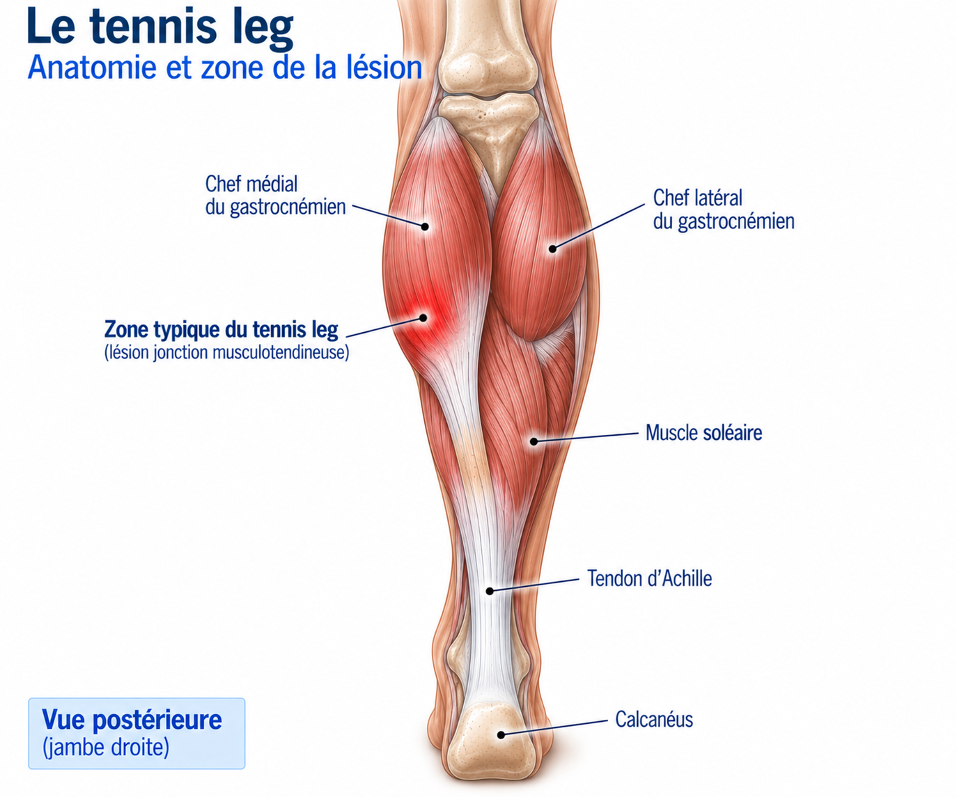
🔍 Click on the image to enlarge it
The medial gastrocnemius
The medial gastrocnemius is the structure most often involved in tennis leg. The vulnerable zone is usually located in its distal part, close to the junction with the soleus aponeurosis, the myotendinous structures and the Achilles tendon complex — a region that has to absorb considerable forces during rapid movements. Modern imaging studies confirm that injuries of the medial gastrocnemius are much more frequent than isolated ruptures of the plantaris.
The role of the triceps surae
The triceps surae is mainly responsible for ankle plantar flexion, propulsion, jumping, running, acceleration and ankle stabilisation; it is also involved in absorbing forces during landing, running and deceleration.
Biomechanics and mechanism
The classic mechanism combines knee extension, ankle dorsiflexion and contraction of the triceps surae: the gastrocnemius is then suddenly stretched while simultaneously trying to produce considerable force, which can cause a muscle or myotendinous injury. This mechanism can occur during an explosive start, a sprint, an acceleration, a change of direction, a jump, a landing or a lateral movement. The typical scenario is an athlete pushing off their foot as the knee straightens: the gastrocnemius is then activated, strongly stretched and subjected to a high eccentric load; when the stress exceeds the tissue’s capacity, an injury can occur.
Epidemiology
Tennis leg can occur in many activities (tennis, padel, running, football, rugby, skiing, racket sports, jumping sports, hiking) and even during everyday activities, in people who do not play sport regularly. It frequently occurs in middle-aged adults, particularly over the age of 40: returning to an explosive activity after a period of low activity is a particularly classic situation.
Causes & risk factors
The injury results from a sudden stretch of the gastrocnemius while it is producing a forceful contraction, typically during an explosive start, a sprint, an acceleration, a change of direction, a jump, a landing or a lateral movement. When the stress exceeds the tissue’s capacity at the precise moment the muscle is both stretched and activated, a muscle or myotendinous injury can occur.
A particularly classic risk factor is returning to an explosive activity after several weeks of low activity or time off sport, without gradually re-exposing the calf to rapid loads — a common situation in middle-aged athletes resuming intense activity (tennis, padel, sprinting) without sufficient preparation.
Symptoms
Patients often describe a very characteristic sensation: “it felt like I’d been kicked in the calf” or “I felt something snap at the back of my calf”. The pain usually comes on suddenly, during exertion or a quick movement, and the person often has to stop their activity immediately.
The main symptoms are sudden calf pain, a snapping sensation or a feeling of having been struck, difficulty walking, pain on plantar flexion, pain when stretching the calf, variable swelling and delayed bruising. The pain is usually medial and posterior, in the middle or lower third of the calf, and may sometimes extend down towards the ankle or the Achilles tendon, or become more diffuse as the haematoma spreads.
Walking may be difficult immediately after the injury (limping, reduced weight-bearing, difficulty rolling through the step, pain on push-off): the ability to walk is an important element in monitoring progress. Contraction of the triceps surae can reproduce the pain, making it particularly difficult to rise onto tiptoe, climb stairs, run, jump or push off the forefoot. Stretching the gastrocnemius (knee extended, ankle in gradual dorsiflexion) can also reproduce the pain — but this stretch should not be forced in the acute phase.
Clinical examination
The examination looks at the location of the pain, tenderness on palpation, swelling, bruising, loss of strength, pain on contraction and stretching, the ability to bear weight and the ability to rise onto tiptoe. The Achilles tendon, ankle, knee and blood vessels must also be examined systematically.
Palpation looks for a precise painful area and may reveal tenderness at the myotendinous junction, a gap in continuity in severe injuries, oedema or a haematoma — its location helping to point towards the medial gastrocnemius rather than the Achilles tendon. The contraction test (resisted plantar flexion) may reproduce the pain and is used to look for reduced strength or asymmetry, comparison with the uninjured side being particularly useful. When pain allows, a tiptoe assessment is tested gradually (both feet, then single leg, then repetitions) to assess the functional recovery of the triceps surae.
Further tests
Ultrasound
First-line examination: visualises the medial and lateral gastrocnemius, the soleus, the gastrocnemius-soleus interface, fluid collections, haematomas and muscle ruptures, with possible comparison with the uninjured side. Can be repeated during follow-up to monitor the reduction of the haematoma, healing and the reorganisation of the fibres.
MRI
More comprehensive analysis: exact location, depth, extent of the injury, myotendinous or soleus involvement, haematoma, associated injuries. Particularly useful when the diagnosis is uncertain, when ultrasound is insufficient, in the case of a deep injury, before a return to high-level sport, or when progress is unusual. A larger injury (large haematoma, extensive myotendinous involvement, significant interfascial separation) may point to a longer recovery, but imaging should not be used on its own to decide on a return to sport: functional recovery takes precedence over the mere disappearance of a radiological abnormality.
Differential diagnoses
Achilles tendon rupture
An essential diagnosis that must always be ruled out: it can also cause sudden pain, a snapping sensation, the feeling of being kicked and difficulty walking. It requires a specific examination (Thompson test, palpation of the tendon, checking for a loss of continuity, assessment of plantar flexion) — confusing it with tennis leg can have serious consequences.
Deep vein thrombosis (DVT)
Can cause calf pain, swelling, warmth, tightness and pain that is sometimes progressive, without the same traumatic mechanism as tennis leg. Should always be considered in the case of atypical pain, significant swelling or thromboembolic risk factors.
Ruptured popliteal cyst
Can cause sudden calf pain and sometimes oedema, and may be mistaken for tennis leg; ultrasound often makes it possible to tell them apart.
Injury to the soleus or lateral gastrocnemius
Rarer forms within the spectrum of musculotendinous injuries of the calf.
Plantaris rupture
Long regarded as the typical tennis leg injury, it is in fact much rarer than injury to the medial gastrocnemius.
Muscle haematoma, stress fracture, compartment syndrome, vascular disease, radiculopathy
To be considered depending on the clinical context when the presentation is atypical.
Classification
Grade 1 — Mild injury
Moderate pain, little or no loss of strength, walking relatively unaffected.
Grade 2 — Moderate injury
Significant pain, limited walking, strength deficit, muscle damage visible on imaging.
Grade 3 — Severe injury
Major rupture, marked loss of strength, large haematoma, great difficulty walking.
Grade 4 — Complete rupture or complex injury
A rare situation requiring specialist assessment.
Treatment
Acute phase (first 48 to 72 hours)
Most cases of tennis leg are treated without surgery. Treatment is based on initial protection, appropriate early weight-bearing and progressive rehabilitation: recent data recommend early mobilisation and structured progression rather than prolonged immobilisation. The goals of the acute phase are to protect the injury, control pain, limit worsening of the haematoma and allow appropriate weight-bearing: relative rest, ice if it brings relief (a symptomatic treatment that does not "repair" the muscle), appropriate compression (without causing numbness, pain or a change in the colour of the foot), elevation, and crutches if walking is too painful — to be phased out gradually as soon as normal walking becomes possible again. Rest should remain relative: prolonged immobilisation is generally not desirable. Anti-inflammatories may be used in certain situations to relieve pain, but they are not considered to speed up healing.
Restoring mobility and isometric contractions
As the acute pain subsides, ankle and knee mobility and normal walking are gradually restored, using controlled movements — avoiding aggressive calf stretching in the early phases. Isometric contractions of the triceps surae are then introduced: isometric plantar flexion (pushing the forefoot against resistance, holding for 20 to 30 seconds, 4 to 5 repetitions), with the intensity increased gradually.
Concentric and eccentric strengthening of the gastrocnemius and soleus
When walking is pain-free or almost pain-free, double-leg heel raises (3 × 10-15) are introduced, then single-leg raises (3 × 8-15), gradually increasing repetitions and load. Eccentric work then becomes an important component of recovery (rising on two legs → slow lowering on one leg), progressing in load, range of motion and speed.
Training should not focus solely on the gastrocnemius: the soleus plays a major role in running, propulsion and force absorption. Bent-knee exercises are introduced gradually (seated calf raise, bent-knee calf press, resisted plantar flexion) — a straight knee loads the gastrocnemius more and a bent knee loads the soleus more, so both positions should be included. As strength returns, the load is increased (loaded standing or seated calf raise, calf press, single-leg work with a dumbbell), then the work becomes single-leg (single-leg raises, long and heavy sets, different ranges of motion) to regain strength and endurance comparable to the uninjured side.
Power, plyometrics and return to running
Once strength is restored, speed must be reintroduced (small hops, jumps on the spot, skipping rope, bounds, horizontal jumps) before returning to sprinting, then progressing through plyometrics: small double-leg hops, repeated double-leg jumps, single-leg jumps, bounds, horizontal jumps, then sport-specific plyometrics.
Running is reintroduced when walking is normal, tiptoe raises are well tolerated, strength is sufficiently restored and dynamic exercises are pain-free or almost pain-free: brisk walking, slow jogging, continuous running, light then fast accelerations, then sprinting.
Complementary treatments
Massage
May improve comfort and reduce the feeling of tightness, as a complement.
Mobilisation
May help to gradually restore mobility.
Compression
Useful in certain phases to control swelling.
Taping
May temporarily improve comfort.
Shockwave therapy
No routine indication in acute tennis leg: it is a musculotendinous injury in the healing phase, where the priority remains mobilisation, progressive strengthening and functional retraining.
PRP (platelet-rich plasma)
Evidence specific to tennis leg is insufficient for it to be a standard treatment; not routinely recommended.
Surgery
Most cases of tennis leg do not require surgery. Surgery may exceptionally be discussed for certain complete ruptures, very extensive injuries, particular complications or the exceptional failure of conservative treatment. Conservative treatment remains the rule.
Recovery time
Healing begins quickly, but full functional recovery takes longer. Depending on severity, recovery can take around 6 to 12 weeks, with considerable variation depending on the injury; recent data report full recovery ranging from approximately 3 to 16 weeks depending on the case, with more severe or more extensive injuries requiring more time.
Before returning to running, the ideal is normal walking, no significant pain, satisfactory ankle mobility, rising onto tiptoe without significant pain, sufficient strength and good tolerance of dynamic exercises. For a full return to sport, a recent clinical review proposes as a practical benchmark an asymmetry in strength, power, endurance and dorsiflexion of less than about 10% compared with the uninjured side.
Prevention
Preventing recurrence relies mainly on regular calf strengthening (working the gastrocnemius and the soleus), single-leg work, power and plyometric work, and gradual exposure to sprinting. The classic situation of several weeks without running followed by a direct return to sprinting must be avoided: the muscle must be gradually re-exposed to rapid loads.
As tennis leg frequently occurs in middle-aged adults — returning to an explosive activity after a period of low activity being a particularly classic situation after the age of 40 — it is worth regularly keeping up calf raises, single-leg work, jumps, running and accelerations, in order to maintain sufficient muscle capacity even during periods of reduced sporting activity.
Frequently asked questions
Is tennis leg a tendinopathy?
Is it a rupture of the plantaris?
Can you walk with tennis leg?
Do I need an MRI?
How long does it take to heal?
When can you return to running?
When can you return to sprinting?
Can you get tennis leg without playing tennis?
Can I cycle?
Is shockwave therapy useful?
Common mistakes
Returning too quickly: pain can subside quickly while muscle capacity remains insufficient.
Staying completely at rest for too long: prolonged immobilisation promotes loss of strength.
Neglecting the soleus: the gastrocnemius is not the only muscle to rehabilitate.
Doing only stretches, when strength and power need to be restored.
Going straight back to sprinting: speed must be reintroduced gradually.
Relying solely on MRI: a persistent abnormality on imaging does not necessarily mean the athlete is not ready to return.
Forgetting about Achilles tendon rupture: any sudden calf pain should prompt an examination of the Achilles tendon.
Forgetting about venous thrombosis: atypical calf pain or swelling should prompt a search for DVT when the context warrants it.
Key points — Institut Basque du Coureur
Tennis leg mainly corresponds to an acute injury of the medial gastrocnemius, often located at its myotendinous junction with the soleus structures. It typically occurs during a sudden movement combining knee extension, ankle dorsiflexion and calf contraction. The classic presentation combines sudden pain, a snapping or kicked sensation, posteromedial calf pain, difficulty walking and pain on plantar flexion. The diagnosis is mainly clinical, with ultrasound or MRI used depending on the context. Two diagnoses in particular must be ruled out: Achilles tendon rupture and deep vein thrombosis. Management is generally conservative: initial protection, then mobility, strengthening, single-leg work, power, plyometrics, running, sprinting and finally sport-specific movements. Functional recovery is generally very good. Tennis leg is not simply calf pain: it is a musculotendinous injury whose healing requires the gradual rebuilding of strength, power and the capacity to absorb stress — the disappearance of pain is not enough to return to sprinting.
- Schwach M, Gaulin B, Vermorel PH, Horteur C, Tourné Y. Tennis leg: Diagnosis and management – A state-of-the-art review. Foot (Edinb). 2026;68:102267.
- Godoy IRB, et al. Imaging in the diagnosis and characterization of tennis leg. Skeletal Radiol. 2026.
- Shah JR, Shah BR, Shah A. Pictorial essay: Ultrasonography in 'tennis leg'. Indian J Radiol Imaging. 2010;20(4):269-273.
- Delgado GJ, Chung CB, Lektrakul N, et al. Tennis leg: clinical US study of 141 patients and anatomic investigation of four cadavers with MR imaging and US. Radiology. 2002.
- Bianchi S, Abdelwahab IF, Mazzola CG, et al. Sonographic evaluation of the gastrocnemius muscle in tennis leg.
- Kwak HS, Lee JM, Lee YS, et al. Diagnosis and follow-up MRI evaluation of tennis leg: New understanding of the pathogenesis and imaging. Injury. 2021.
- Ryu JA, Lee JH. Ultrasound evaluation of calf injuries.
- Thompson JM, et al. Medial Gastrocnemius Strain: Clinical Aspects and Algorithmic Approach. 2023.
- Pollock N, James SLJ, Lee JC, Chakraverty R. British athletics muscle injury classification and grading system. Br J Sports Med. 2014.
- Mueller-Wohlfahrt HW, Haensel R, Mithoefer K, et al. Terminology and classification of muscle injuries in sport: the Munich consensus statement. Br J Sports Med. 2013.
- Askling CM, Tengvar M, Saartok T, Thorstensson A. Acute first-time hamstring strains during high-speed running: a longitudinal study including clinical and MRI findings.
- Silder A, Heiderscheit BC, Thelen DG, Enright T, Tuite MJ. MR observations of long-term muscle recovery following hamstring strain injury.
Similar injuries
Compartment syndrome
Increased muscle pressure during exercise that can compromise blood flow and nerve function.
Popliteal artery entrapment syndrome
A rare but serious vascular compression of the popliteal artery that causes exercise-induced pain.
Tibial stress fracture
A tibial micro-fracture caused by repeated stress, requiring a mandatory break from running to allow the bone to heal.