
Definition
Tibialis posterior tendinopathy is a condition of overload and progressive dysfunction of the tendon of the tibialis posterior muscle. It initially presents as pain, most often located on the medial side of the ankle or foot, sometimes associated with mild swelling along the course of the tendon.
The tibialis posterior plays an essential role in supporting the medial arch of the foot and in controlling foot pronation. When its capacity to produce and transmit sufficient force decreases, the foot may gradually lose part of its dynamic support.
This condition is part of a continuum ranging from a painful tendinopathy without significant deformity to a progressive deformity of the foot with collapse of the arch. The historical term “posterior tibial tendon dysfunction” (PTTD) is still widely used in the literature; however, more recent classifications place greater emphasis on the overall deformity of the foot and use the term Progressive Collapsing Foot Deformity (PCFD).
In advanced forms, the damage is no longer confined to the tendon: it gradually extends to the ligaments, joints and bones. Management must therefore be tailored to the stage of the disease, which is why early recognition and treatment are important.
Anatomy
The tibialis posterior is a deep muscle of the posterior compartment of the lower leg. It originates from the posterior surface of the tibia, the posterior surface of the fibula and the interosseous membrane that connects them.
Its tendon runs down behind the medial malleolus before heading towards the medial side of the foot. It inserts mainly on the navicular, with expansions to the cuneiforms, the cuboid and the bases of some metatarsals — an anatomy that explains its major role in supporting the arch of the foot.
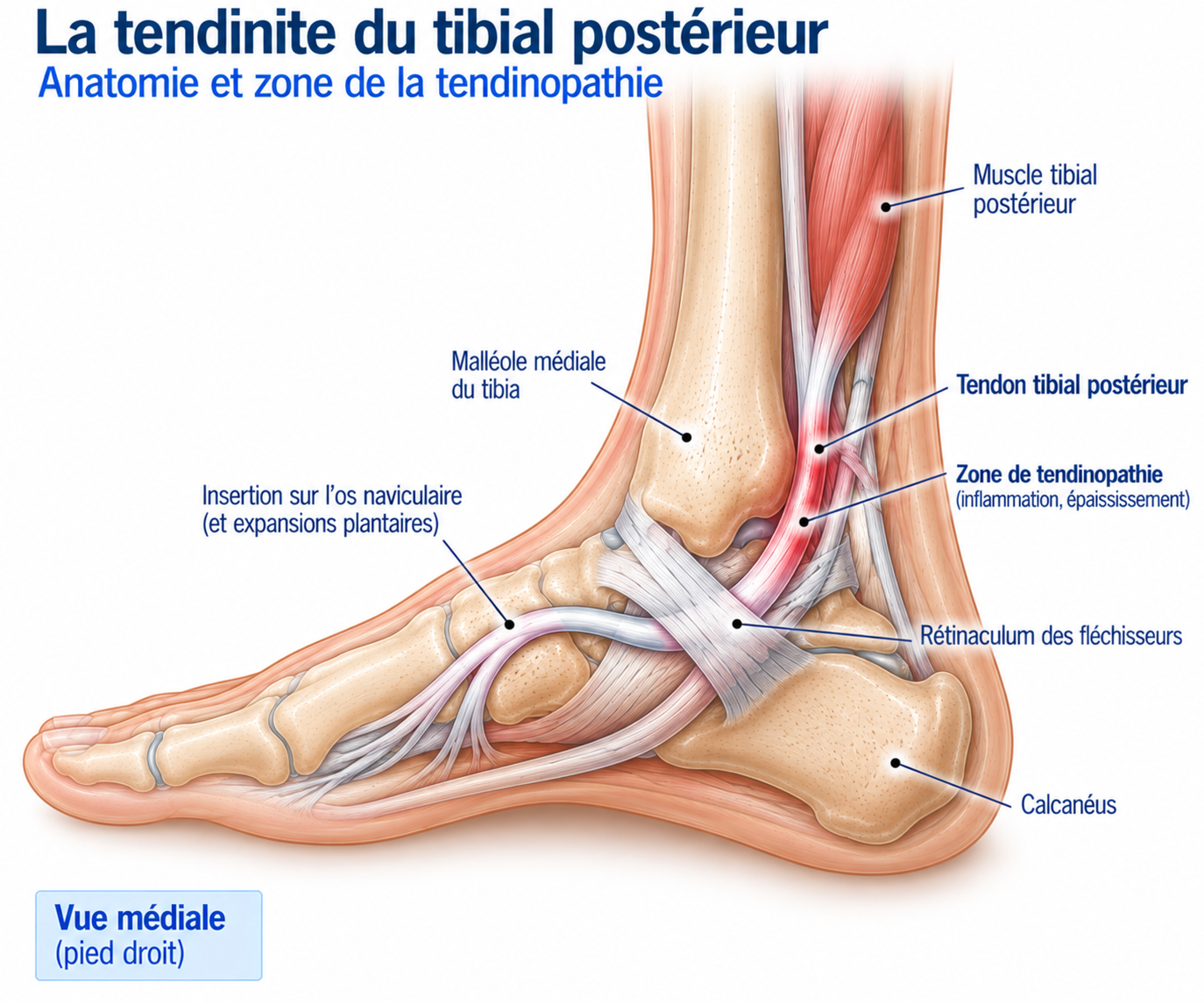
🔍 Click on the image to enlarge it
The tendon never works alone: it acts in close association with the plantar calcaneonavicular ligament (“spring ligament”), the plantar fascia, the deltoid ligament, the intrinsic foot muscles, the tibialis anterior and the fibular muscles. It is thus one of the main dynamic stabilisers of the medial arch.
Biomechanics of the tibialis posterior
The tibialis posterior contributes to foot supination, inversion, plantar flexion, hindfoot stabilisation and support of the medial arch. During walking and running, the foot must be mobile enough to absorb stress at foot strike, then rigid enough to transmit forces efficiently during push-off: the tibialis posterior contributes to this transformation by controlling pronation.
During the stance phase, a certain amount of pronation is physiological; the tibialis posterior slows its range and thus works as a true active muscular brake. As push-off approaches, the foot must instead become more rigid: the muscle contributes to foot supination and arch stabilisation, turning the foot into an efficient propulsion lever.
Role in supporting the arch of the foot
Together with the spring ligament, the plantar fascia and the other foot muscles, the tibialis posterior forms a dynamic and passive support system for the arch. When the tendon becomes painful or loses mechanical capacity, a chain of events can gradually develop: reduced tibialis posterior strength → increased pronation → lowering of the arch → increased ligament loading → progressive foot deformity.
This progression is not systematic, however. Early tendinopathy can remain stable for several years without progressing to significant deformity. The aim of early management is precisely to prevent the loss of tendon capacity from becoming great enough to cause this deformity.
Epidemiology
Tibialis posterior tendinopathy is particularly common in middle-aged and older adults. It is less common in young athletes than Achilles or patellar tendinopathy, but it can nevertheless occur in athletes exposed to repeated running or walking stresses.
Several factors are associated with this condition: age, excess weight, high blood pressure, diabetes, dyslipidaemia, flat feet, a history of ankle injury and a substantial increase in activity load.
The true prevalence varies depending on the populations studied and the diagnostic criteria used; it is probably underdiagnosed, particularly in the early stage of the disease.
Causes & risk factors
Tibialis posterior tendinopathy is a multifactorial condition, most often resulting from a combination of several mechanical, muscular, metabolic and age-related factors.
Mechanical factors
Several factors increase the stress placed on the tendon: flat feet, overpronation, hindfoot valgus, a sudden increase in activity, prolonged walking, running, prolonged standing at work and excess weight.
Having flat feet does not, however, mean that tendinopathy will necessarily develop: the foot can function perfectly well with a low arch if muscular and ligamentous capacity remains sufficient.
Flat feet: should you be worried?
Flat feet are not necessarily pathological. They become a concern when they are painful, progressively worsening, associated with a loss of function or with an inability to perform a single-leg heel raise. A low but stable and asymptomatic arch does not systematically require treatment: the key question is not “does the patient have flat feet?” but “is this flat foot symptomatic, functionally insufficient or progressively deforming?”
Factors related to muscle strength
Reduced strength of the tibialis posterior and triceps surae reduces the capacity of the lower limb to control the stresses placed on the foot. The intrinsic foot muscles also play an important role in stabilising the arch.
Metabolic factors
Several conditions are associated with an increased risk: diabetes, high blood pressure, obesity and dyslipidaemia. A combination of several of these metabolic factors appears to be particularly unfavourable.
Age-related factors
The tendon’s capacity to adapt to mechanical stress gradually decreases with age, which explains why this condition is much more common in middle-aged and older adults.
Symptoms
The main symptom is pain on the medial side of the ankle or foot, generally following the course of the tendon: behind the medial malleolus, along the medial border of the foot, down to its insertion on the navicular.
At first, pain appears only during activities that place heavy demands on the tendon: prolonged walking, running, climbing stairs, hiking or prolonged standing. Swelling along the tendon may also be observed. As the condition progresses, pain may appear during everyday activities.
The gradual loss of tibialis posterior capacity may also show up as difficulty standing on tiptoe on one foot, foot fatigue, a feeling that the arch is collapsing, a gradual increase in pronation or difficulty with prolonged walking. A particularly telling sign is the onset of difficulty performing a single-leg heel raise: the patient may at first manage it with pain or with reduced heel height, then become unable to perform the movement properly in more advanced forms.
Clinical examination
The examination should assess the state of the tendon, muscle strength, foot alignment, mobility and function all at once. Inspection looks for reduced medial arch height, hindfoot valgus, forefoot abduction, a change in the axis of the calcaneus and asymmetry between the two feet. Viewed from behind, increased visibility of the lateral toes on the affected side (“too many toes sign”) may indicate forefoot abduction; this sign is classically looked for in advanced forms.
The Single Leg Heel Rise Test is one of the most important functional tests: the patient raises the heel while standing on one foot, which should normally be accompanied by progressive inversion of the calcaneus. Pain, reduced heel height, rapid fatigue, an inability to repeat the movement or a lack of calcaneal inversion suggest tibialis posterior dysfunction. This test simultaneously assesses triceps surae strength, tibialis posterior function and the capacity of the arch to withstand stress; it is also an excellent tool for monitoring rehabilitation.
Assessment of ankle and foot mobility looks for limited dorsiflexion or supination, hindfoot stiffness, or limited mobility of the midtarsal joints and the first ray. An essential distinction must be made between a flexible deformity (still passively correctable) and a rigid deformity (not correctable), which is of major importance for prognosis and treatment strategy. Finally, the muscle assessment looks for weakness of the tibialis posterior, triceps surae, intrinsic foot muscles, tibialis anterior and fibular muscles — ideally assessed under load, in a functional situation, which is more informative than a simple isolated test.
Further tests
Ultrasound
Examines the thickness and structure of the tendon, looks for fibrillar abnormalities, splits or a rupture, as well as vascularisation and movement of the tendon during a dynamic examination. It is the most widely used first-line imaging modality in clinical practice.
MRI
Examines the tibialis posterior tendon, the spring ligament, the joints, the bony structures and the surrounding soft tissues simultaneously. Particularly useful when a rupture or associated damage is suspected.
Weight-bearing X-rays of the foot
Particularly useful when a deformity is suspected: they make it possible to assess arch height, hindfoot alignment, forefoot abduction and any associated arthropathy. A foot that looks normal when not bearing weight can show significant deformity under load.
Differential diagnoses
Stress fracture
Pain more localised over a specific bony landmark, to be distinguished from pain following the course of the tendon, which is characteristic of tibialis posterior tendinopathy.
Medial ankle sprain / deltoid ligament injury
An acute traumatic event can usually be identified, unlike the gradual onset typical of tendinopathy.
Spring ligament (plantar calcaneonavicular ligament) injury
Often associated with or secondary to tibialis posterior insufficiency; when present, it contributes to collapse of the arch.
Fibular or flexor hallucis tendinopathy
Pain that is more lateral (fibulars) or posterior plantar (flexor hallucis), to be distinguished by precise palpation along the course of the tendon.
Tarsal tunnel syndrome
Pain associated with paraesthesia or neurological involvement of the tibial nerve, which are absent in isolated tibialis posterior tendinopathy.
Subtalar, talonavicular or midtarsal osteoarthritis, medial ankle impingement
Pain that is more articular than tendinous, often associated with stiffness; more common in advanced forms with rigid deformity.
Classification
Stage 1
The tendon is painful but the shape of the foot is broadly preserved, with no significant deformity. Strength may be reduced but function remains relatively preserved.
Stage 2
The tendon becomes insufficient and a flexible deformity appears: lowering of the arch, hindfoot valgus, forefoot abduction. The deformity remains passively correctable.
Stage 3
The deformity becomes more pronounced and progressively rigid, sometimes with degenerative joint changes. Passive correction becomes limited.
Stage 4
The foot deformity is accompanied by involvement of the ankle joint, particularly the deltoid ligament complex. This is an advanced form requiring specialist management.
Treatment
Adjusting training load
In painful forms, activities that cause significant overload should be temporarily reduced: running, hiking, long walks, prolonged standing or repeated jumping. Complete rest, however, is generally not necessary.
Better-tolerated activities can be continued, such as cycling, swimming or other low-impact cardiovascular exercise, in order to maintain fitness while temporarily reducing the stress placed on the tendon.
Physiotherapy rehabilitation (progressive, phased approach)
Rehabilitation is the reference treatment and progresses in successive stages, adapted to the tendon's tolerance.
Phase 1 — symptom control: gradual reintroduction of tendon loading through isometric contractions in inversion, plantar flexion against resistance, double-leg exercises, active ankle mobilisation and work on the intrinsic foot muscles.
Phase 2 — strengthening the tibialis posterior: more functional work combining inversion against resistance, combined plantar flexion + inversion, then heel raises, first on both legs and then assisted on one leg, progressing from bilateral to unilateral and then to added load. Available data suggest a benefit from eccentric strengthening when combined with orthoses and stretching, although the level of evidence is less robust than for other tendinopathies.
Phase 3 — strengthening the arch support system: the tibialis posterior is not strengthened in isolation; the programme also targets the soleus, gastrocnemius, tibialis anterior, intrinsic foot muscles, fibular muscles and hip muscles. A Short Foot exercise (actively shortening the foot without clenching the toes) and work on controlling pronation under load complete this phase.
Phase 4 — restoring dynamic function: once strength has been restored, the work becomes more functional — single-leg balance, exercises on unstable surfaces, walking on tiptoe, climbing stairs, lunges, squats, single-leg exercises, then jumps if the sporting level requires it.
The ability to perform single-leg heel raises is an excellent indicator of progress: one published protocol for early forms used the completion of 50 single-leg heel raises with minimal or no pain as its criterion for success. This figure should not, however, be considered a universal threshold applicable to all patients.
Gradual return to running
In athletes, the return to running must be gradual and is only considered once walking is well tolerated, strengthening exercises are performed without a significant reaction, single-leg control is satisfactory, heel raises are performed correctly, and pain and any swelling remain stable the following day.
The return can follow a progression such as: walking → slow running → continuous running → increased volume → increased speed → hills → interval training, never increasing volume, speed and elevation gain at the same time.
Complementary treatments
Foot orthoses
Particularly worthwhile in cases of flexible deformity of the arch: they support the arch, reduce stress on the tendon and limit excessive pronation. However, they should not replace muscle strengthening — the principle remains: orthosis = reduced stress, rehabilitation = increased capacity.
Suitable footwear
A stable shoe, with good midfoot support and a sufficiently stiff sole, can reduce some of the stress on the foot. Abrupt changes of footwear should be avoided.
Temporary immobilisation
May be considered in some very painful forms or during a marked inflammatory phase, but must always be followed by rehabilitation to avoid deconditioning.
Anti-inflammatories
May be of occasional symptomatic benefit, but they do not correct the mechanical cause of the condition.
Extracorporeal shockwave therapy
The available data are much more limited than for other tendinopathies: they are not the reference treatment.
Corticosteroid injections
Injections directly into the tendon should be avoided because of the risk of weakening it. An intra-articular injection may possibly be discussed in cases of associated symptomatic arthropathy, but it does not treat the tendon dysfunction itself.
Surgery
The surgical strategy depends mainly on the stage of the disease. In early forms, conservative treatment remains the preferred option. In forms with flexible deformity, reconstructive surgery may be considered when several months of well-conducted conservative treatment have not produced sufficient improvement. In advanced forms with rigid deformity or joint involvement, surgery may become necessary.
Depending on the situation, procedures may combine tendon repair or debridement, tendon transfer, calcaneal osteotomy, correction of midfoot deformity, ligament reconstruction or, in advanced arthritic forms, arthrodesis. The strategy therefore depends far more on the stage of the deformity than on the mere presence of tendinopathy.
A specialist opinion is particularly recommended in cases of persistent pain despite properly conducted management, suspected tendon rupture, inability to perform a single-leg heel raise, progressive or rigid deformity, newly developed flat foot in an adult, associated joint pain or suspected spring ligament involvement.
Recovery time
Recovery time depends heavily on the stage of the disease. Early tendinopathy can begin to improve within a few weeks with proper load adjustment and progressive rehabilitation. More advanced forms, particularly when a foot deformity is already present, may require several months of management.
The earlier management begins, the more it is possible to favour a conservative strategy and avoid progression to a structural deformity of the foot.
Prevention
Prevention relies on maintaining good functional capacity of the foot: regular strengthening of the tibialis posterior and triceps surae, work on the intrinsic foot muscles, balance training, pronation control and maintaining good ankle mobility.
In athletes, periods of returning to sport after a break should be particularly gradual, with a carefully planned increase in training load. In people with flat feet, monitoring for the onset of medial pain or a gradual decrease in the ability to stand on tiptoe makes it possible to intervene early, before a deformity sets in.
Frequently asked questions
Can you run with tibialis posterior tendinopathy?
Do flat feet mean that the tibialis posterior is damaged?
Are orthopaedic insoles essential?
Can the tendon be strengthened despite the pain?
Are eccentric exercises effective?
Do I need an MRI?
Is an X-ray needed?
Can tendinopathy progress to flat feet?
Can surgery be avoided?
How long does recovery take?
Common mistakes
Regarding every flat foot as pathological.
Waiting until a significant deformity appears before seeing a doctor.
Treating only the pain without correcting the mechanical cause.
Relying solely on orthopaedic insoles without any strengthening work.
Stopping all activity completely for several weeks.
Returning abruptly to activity after symptoms improve.
Neglecting strengthening of the triceps surae and the intrinsic foot muscles.
Multiplying imaging tests without correlating them with the clinical examination.
Key points — Institut Basque du Coureur
Tibialis posterior tendinopathy should not be regarded as simple tendon pain in the ankle. The tibialis posterior plays an essential role in the dynamic support of the arch and the control of pronation: a gradual decrease in its capacity can lead to an acquired flat foot and, in advanced forms, to a true foot deformity. Management is based early on load adjustment and progressive strengthening of the tibialis posterior and the entire muscular system of the foot, combined with an orthosis if necessary. The earlier management begins, the more it is possible to preserve function and avoid progression to a structural deformity.
- Ross MH, Smith MD, Mellor R, Vicenzino B. Exercise for posterior tibial tendon dysfunction: a systematic review of randomised clinical trials and clinical guidelines. BMJ Open Sport Exerc Med. 2018;4:e000430.
- Bowring B, Chockalingam N. A clinical guideline for the conservative management of tibialis posterior tendon dysfunction. The Foot. 2009;19(4):211-217.
- Alvarez RG, Marini A, Schmitt C, Saltzman CL. Stage I and II posterior tibial tendon dysfunction treated by a structured nonoperative management protocol: an orthosis and exercise program. Foot Ankle Int. 2006.
- American College of Foot and Ankle Surgeons. Clinical Consensus Statement: Appropriate Clinical Management of Adult-Acquired Flatfoot Deformity. J Foot Ankle Surg. 2020.
- Kohls-Gatzoulis J, Woods B, Angel JC, Singh D. The prevalence of symptomatic posterior tibial tendon dysfunction in women over the age of 40 in England. Foot Ankle Surg.
- Kulig K, Reischl SF, Pomrantz AB, et al. Nonsurgical management of posterior tibial tendon dysfunction with orthoses and resistive exercise: a randomized controlled trial.
- Kulig K, Burnfield JM, Requejo SM, et al. Posterior tibial and tibialis anterior muscle activity in persons with and without posterior tibial tendon dysfunction.
- Myerson MS. Adult acquired flatfoot deformity: treatment of dysfunction of the posterior tibial tendon. Instr Course Lect.
- Johnson KA, Strom DE. Tibialis posterior tendon dysfunction. Clin Orthop Relat Res. 1989.
- Bluman EM, Myerson MS, Symms CL. Posterior tibial tendon rupture: a refined classification system. Foot Ankle Clin.
- 2024 UK survey of non-surgical management of posterior tibial tendon dysfunction. Journal of Foot and Ankle Research.
Physiotherapy protocol for this injury
Découvrez notre protocole de rééducation détaillé pour tibialis posterior tendinopathy : exercices adaptés, progression structurée et modalités de reprise.
Similar injuries
Achilles tendinopathy
Inflammation of the Achilles tendon causing pain and stiffness at the heel, the most common injury in runners.
Plantar plate injury
Damage to the fibrous plate that stabilises the toes, causing pain under the forefoot and a feeling of walking on a pebble.
Morton's neuroma
Compression of an interdigital nerve in the forefoot, causing pain and numbness.