
Definition
Iliotibial band syndrome, also called ITB syndrome or Iliotibial Band Syndrome (ITBS), is an overuse condition that causes pain on the outer side of the knee. It is one of the main causes of lateral knee pain in runners, but it also affects cyclists and athletes whose sports involve repeated knee flexion-extension movements. Its incidence is estimated at between 5 and 14% of running-related injuries.
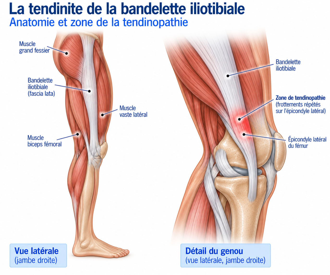
The historical term “iliotibial band friction syndrome” is based on the idea that the band rubs repeatedly against the lateral femoral condyle. However, current biomechanical data suggest that the mechanism is probably more complex: the iliotibial band is a relatively stiff fibrous structure which, during repeated flexion-extension movements, would mainly increase the compression of the tissues lying between it and the lateral femoral condyle, particularly at around 20 to 30° of knee flexion.
The syndrome is therefore probably more a matter of mechanical and compressive overload of the lateral knee tissues than a simple friction phenomenon. It is generally linked to an increase in training load, combined with biomechanical and neuromuscular factors that can increase the stresses on the lateral compartment of the knee.
Anatomy
The iliotibial band is a longitudinal fibrous structure on the lateral side of the thigh. It is the thickened part of the fascia lata and receives fibres from the tensor fasciae latae and the gluteus maximus. It runs down the outer side of the thigh and inserts on the anterolateral part of the tibia, at Gerdy’s tubercle, contributing to the lateral stability of the hip and knee.
At the knee, it is closely related to the lateral femoral condyle, the lateral retinaculum, the joint capsule, the vastus lateralis, and the fatty and connective tissue structures beneath the band. This anatomy explains why a repeated increase in compression in this region can become painful.
🔍 Click on the image to enlarge it
Biomechanics of the iliotibial band
The iliotibial band helps control the position of the lower limb in the frontal and transverse planes: it contributes to lateral knee stability, to the transmission of forces between the pelvis and the lower limb, to hip stabilisation and to limiting certain excessive movements of the lower limb.
During running, the knee passes several times per cycle through a range of flexion in which the stresses on the lateral tissues can be considerable. The repetition of thousands of cycles can become a problem when the load increases faster than the tissues’ capacity to adapt. However, the mechanism is not purely local: the biomechanics of the pelvis, hip, knee and foot all play a part in how the stresses are distributed.
The role of the hip and pelvis
Hip control plays an important role in running biomechanics. Reduced frontal-plane control of the pelvis or an increase in certain hip movements can alter the relative position of the knee and increase the stresses on the lateral structures. In some studies, runners with iliotibial band syndrome show differences in hip, knee and ankle kinematics compared with asymptomatic runners.
However, the findings in the literature remain heterogeneous: hip abductor weakness may be a contributing factor in some runners, but on its own it is not a universal explanation for the syndrome.
Epidemiology
Iliotibial band syndrome is particularly common in distance runners. It accounts for a significant proportion of running-related injuries and is one of the most common causes of pain in the lateral compartment of the knee. It also affects cyclists, triathletes, hikers, endurance athletes and players of some team sports.
In runners, it frequently occurs during periods of increasing training load. Preparing for a half marathon, marathon or trail race can be a period of particular risk when volume or elevation gain increases rapidly.
Causes & risk factors
Iliotibial band syndrome is multifactorial. It is generally linked to a combination of high mechanical load, insufficient recovery, biomechanical factors and insufficient muscle capacity of the pelvic chain.
Training load and terrain
The main trigger is often a recent increase in load: more mileage, more frequent sessions, a return after a break, higher speed, the introduction of interval training, a large increase in elevation gain, more descents or a sudden increase in the length of runs. Downhill running is particularly likely to increase the repeated stresses on the knee.
Terrain can also influence the stresses: prolonged running on a camber, on strongly cambered roads, on technical trails or downhill can alter lower limb kinematics. Repeated changes of direction can also increase lateral stresses.
Neuromuscular factors
Control of the pelvis and hip is an important part of the assessment: weakness of the hip abductors, deficits in the gluteus medius or gluteus maximus, reduced pelvic control, poor single-leg stability or insufficient knee control during running. Hip abductor strengthening is one of the most frequently studied management strategies: a systematic review published in 2024 found it to be a strategy common to the various studies, with improvements in pain and function over periods of 2 to 8 weeks. However, the syndrome should not be reduced to simple “gluteus medius weakness”.
Biomechanical factors
Biomechanical studies have found some differences between runners with and without iliotibial band syndrome in hip kinematics, lower limb rotation, knee movements and foot kinematics. One systematic review found differences in lower limb kinematics in runners with iliotibial band syndrome, but the results vary from study to study: there is therefore no single biomechanical profile of the affected runner, and the assessment must be individualised.
Symptoms
The main symptom is pain on the outer side of the knee, usually very well localised around the lateral femoral condyle. Runners often describe pain at a precise point, a burning sensation that is sometimes very sharp, appearing after a certain amount of time running.
A particularly characteristic feature is the predictable nature of symptom onset — for example, “the pain always starts after 25 minutes of running”. At first, the pain appears only during running and usually disappears quickly after stopping: the athlete may be completely symptom-free at rest, when walking and in daily life. As the condition progresses, the time before pain appears gradually shortens; in more severe forms, it can persist after the session and cause difficulty on stairs or with some daily activities.
Clinical examination
The diagnosis is mainly clinical. The examination looks for precise pain over the lateral femoral condyle, brought on by repeated knee flexion-extension, by running or by single-leg movements, as well as impaired pelvic control, weakness of the abductors or gluteus maximus and any abnormalities in running mechanics. Palpation of the lateral femoral condyle usually reproduces the pain precisely.
Two clinical tests are classically used: Noble’s test (pressure applied to the lateral part of the femoral condyle while the knee is gradually flexed, with reproduction of the pain being suggestive) and Ober’s test (looking for limited hip adduction with the lower limb extended). Its diagnostic value should, however, be interpreted with caution: a positive Ober’s test does not necessarily mean that the band is “too short” or that it needs to be stretched, as modern biomechanical data call into question the idea that simple stretching can significantly change its mechanical length.
Functional assessment completes the examination: the single-leg squat allows observation of pelvic stability, hip control, knee position and foot control; the step-down test (controlled stepping down from a step) looks at pelvic control, hip adduction and movement quality; running analysis can look at cadence, stride length, pelvic position, vertical oscillation and any asymmetries.
Further tests
Ultrasound
May show thickening of the tissues around the band, abnormalities of the underlying tissues or fluid in a bursa, if present. Mainly useful for looking for another condition of the lateral compartment of the knee.
MRI
May reveal oedema or high signal in the tissues beneath the band, thickening of the band itself, bursitis or associated lesions (meniscus, cartilage). Particularly useful when another cause of lateral knee pain is suspected.
Differential diagnoses
Lateral meniscus injury
Pain often posterolateral or over the joint line, sometimes accompanied by locking, effusion or instability — signs absent in iliotibial band syndrome.
Lateral tibiofemoral osteoarthritis
Tends to affect older people, with diffuse mechanical pain in the lateral compartment and morning stiffness, without the predictable pattern linked to a specific running time.
Patellofemoral pain syndrome
Pain more anterior or peripatellar, made worse by prolonged sitting and stairs, unlike the pinpoint pain over the lateral femoral condyle in iliotibial band syndrome.
Biceps femoris or popliteus tendinopathy
Pain more posterolateral, reproduced by specifically loading these tendons rather than by repeated knee flexion-extension alone.
Lateral collateral ligament injury / knee sprain
A history of trauma found on questioning, sometimes with varus laxity on examination — absent in an overuse condition.
Stress fracture, osteochondral lesion or fabella syndrome
Pain often deeper, sometimes at night, made worse by simple weight-bearing; imaging (MRI) is needed at the slightest doubt.
Referred hip pain or radicular pain
Examination of the lumbar spine and hip is expected to be normal in iliotibial band syndrome; these causes should be systematically looked for if the presentation is atypical.
Classification
Stage 1
Pain appears only after the session. Daily life remains completely symptom-free.
Stage 2
Pain appears during running but after a relatively long time. The athlete can usually continue with modified activity.
Stage 3
Pain appears quickly during running and limits the length of sessions. A significant change in load becomes necessary.
Stage 4
Pain appears during daily activities or persists after exercise. The diagnosis should be reassessed and differential diagnoses systematically looked for.
Treatment
Load adjustment and relative rest
The first step is to temporarily reduce the load responsible for the symptoms, without necessarily stopping all physical activity. Cycling, swimming and strength training can usually be continued as long as they remain pain-free, which helps maintain cardiovascular fitness during the desensitisation phase.
A practical strategy is to identify the tolerance window: the running time before pain appears. If it usually appears after 25 minutes, running can be resumed for a clearly shorter time — for example 15 to 20 minutes — and then increased gradually as long as the pain remains absent or mild, does not increase during the session and disappears quickly after stopping. This approach avoids the classic pattern: complete rest, pain disappears, return at the previous level, then recurrence.
Physiotherapy rehabilitation (first-line treatment)
Rehabilitation is the cornerstone of treatment. It does not simply aim to "loosen the band", but above all to reduce the irritating stresses and then gradually restore the lower limb's capacity to withstand the stresses of running. The available data generally support conservative management, but the literature remains heterogeneous and does not allow a single protocol superior to all others to be defined.
The first phase aims to restore motor control of the lower limb: hip abductor activation, strengthening of the gluteus medius and gluteus maximus, trunk work, pelvic control and single-leg balance, initially performed in low-stress positions.
Strengthening then progresses to more demanding hip exercises (hip abduction, hip hike, side plank, monster walk, single-leg squat), the aim being to restore the functional capacity of the whole pelvic chain rather than to target a single muscle. A third phase integrates this strength into more sport-specific movements (lunges, step-downs, stair climbing, jumps, bounds, multidirectional exercises), requiring pelvic and knee control at increasing speeds.
Finally, a last phase gradually reintroduces the specific stresses of running (easy running, accelerations, changes of pace, hills, descents, interval training), ideally changing only one variable at a time: volume, then frequency, then speed, then elevation gain. Gait analysis (cadence, stride length, pelvic control, knee position) can be useful when mechanical factors clearly contribute to the symptoms, but no change should be applied systematically to all patients.
Gradual return to running
A return to running can be considered when walking is pain-free, single-leg exercises and strengthening are well tolerated, and pelvic and knee control is satisfactory. It often begins with alternating walking and running (for example 2 minutes of walking for 1 minute of running), for a total of about 20 to 30 minutes depending on the patient's level, before gradually increasing the proportion of running.
Progression is considered when the pain remains absent or mild, does not worsen during the session, disappears quickly after stopping and does not lead to a flare-up the next day. A full return to sport requires no pain in daily life, satisfactory abductor and gluteus maximus strength, good motor control (single-leg squat, step-down) and tolerance of continuous running including accelerations, changes of pace and elevation gain.
Complementary treatments
Iliotibial band stretching
The band is a very strong fibrous structure; the available data suggest that conventional stretching has limited capacity to change its length or mechanical properties in a lasting way. It can improve overall hip mobility but should not be the main treatment.
Manual therapy and massage
Can be used as a complementary treatment to address restricted hip mobility or tension in the tensor fasciae latae, with possible short-term symptom relief. Should not replace strengthening and load modification.
Extracorporeal shockwave therapy
May be offered as a complementary treatment in some resistant forms when properly conducted active management has not brought sufficient improvement. The available data remain limited, and they should not replace rehabilitation.
Corticosteroid injection
Can reduce pain quickly in some very painful situations, but does not correct the mechanical factors responsible. It should be seen as an occasional option rather than a long-term treatment; the literature is limited and of variable quality.
Non-steroidal anti-inflammatory drugs (NSAIDs)
May be used for a short period in some painful situations, but they are not the main treatment: the aim remains to correct the mechanical overload and restore functional capacity.
Surgery
Surgery is rarely necessary. It may be discussed in chronic resistant forms after failure of properly conducted conservative management. Historical procedures include release of the band, resection of a pathological portion, bursectomy, or arthroscopic or open procedures depending on the situation.
The reported results are generally favourable in surgical series — one recent systematic review reported return-to-sport rates of 81 to 100% — but these data should be interpreted with caution because of the heterogeneity of the studies and the low level of evidence. Surgery should therefore only be considered after failure of properly conducted conservative treatment.
Recovery time
Recovery time depends mainly on how long the symptoms have been present, the sporting load, how quickly training is adjusted, muscle deficits, biomechanical factors and adherence to rehabilitation. Recent forms can often improve within a few weeks, whereas chronic forms may require several months.
In studies of conservative treatment, improvements in pain and function have been observed over periods ranging from 2 to 8 weeks, but the protocols remain very heterogeneous. A full return to the previous level of sport can take longer than the mere disappearance of pain.
Prevention
Prevention is mainly based on good training load management: increasing mileage gradually, limiting sudden increases in elevation gain, introducing interval training gradually and varying terrain gradually.
Maintaining good hip abductor strength and good pelvic control, and avoiding sudden changes in shoes or running technique, also help limit the risk. In runners who have already had iliotibial band syndrome, regular strengthening of the pelvic chain can be built into the training programme.
Frequently asked questions
Can you keep running with iliotibial band syndrome?
Is it really friction of the band?
Should the iliotibial band be stretched?
Is the gluteus medius always to blame?
Are insoles necessary?
Do I need an MRI?
Is shockwave therapy effective?
Can I cycle?
How long does recovery take?
How can I prevent a recurrence?
Common mistakes
Stopping all activity completely for several weeks.
Going straight back to the previous training level.
Increasing mileage and elevation gain at the same time.
Focusing solely on stretching the band.
Relying only on massage, without muscle strengthening.
Considering gluteus medius weakness to be the sole cause of the syndrome.
Abruptly changing running technique.
Going back to downhill running too quickly.
Neglecting overall strengthening of the lower limb.
Automatically attributing any pain on the outer side of the knee to iliotibial band syndrome, without considering a differential diagnosis.
Key points — Institut Basque du Coureur
Iliotibial band syndrome is an overuse pain in the lateral compartment of the knee, particularly common in runners. Its mechanism probably cannot be reduced to simple friction of the iliotibial band: repeated compression of the tissues beneath it, combined with running load and certain biomechanical factors, probably plays a major role. Treatment is based above all on temporary load adjustment, strengthening of the pelvic chain and lower limb control, followed by a gradual return to running. Stretching or passive treatments can be used as adjuncts, but they are not the core treatment.
- Sanchez-Alvarado A, Bokil C, Cassel M, Engel T. Effects of conservative treatment strategies for iliotibial band syndrome on pain and function in runners: a systematic review. Front Sports Act Living. 2024;6:1386456.
- van der Worp MP, van der Horst N, de Wijer A, Backx FJG, Nijhuis-van der Sanden MWG. Iliotibial band syndrome in runners: a systematic review. Sports Med. 2012;42(11):969-992.
- Friede MC, Innerhofer G, Fink C, Alegre LM, Csapo R. Conservative treatment of iliotibial band syndrome in runners: Are we targeting the right goals? Phys Ther Sport. 2022;54:44-52.
- Nguyen AP, Detrembleur C, Van Cant J. Conservative treatment for iliotibial band syndrome: Are we facing a research gap? A scoping review of 98 studies with clinical perspectives. Phys Ther Sport. 2023;62:25-31.
- Louw M, Deary C. The biomechanical variables involved in the aetiology of iliotibial band syndrome in distance runners: a systematic review. Phys Ther Sport. 2014;15(1):64-75.
- Foch E, Brindle RA, Pohl MB. Lower extremity kinematics during running and hip abductor strength in iliotibial band syndrome: a systematic review and meta-analysis. Gait Posture. 2023;101:73-81.
- Baker RL, Souza RB, Fredericson M. Iliotibial band syndrome in runners: biomechanical implications and conservative management. PM&R.
- Fredericson M, Wolf C. Iliotibial band syndrome in runners: innovations in treatment. Sports Med. 2005.
- Ellis R, Hing W, Reid D. Iliotibial band friction syndrome — a systematic review. Man Ther. 2007;12(3):200-208.
- Barton CJ, Bonanno D, Carr J, et al. Running retraining to treat lower limb injuries: a mixed-methods study of current evidence synthesised with expert opinion. Br J Sports Med.
Similar injuries
Patellar tendinopathy
Overuse tendinopathy of the patellar tendon causing pain below the kneecap, common in runners and jumping athletes.
Knee osteoarthritis
Osteoarthritis of the knee characterised by progressive wear of the cartilage.
Pes anserinus tendinopathy
Inflammation of the tendons that insert on the inner side of the tibia (sartorius, gracilis, semitendinosus), causing pain on the inner side of the knee.